
• O1 Network
    • Articles
    • For Residents
    • For Medical Students
    • PORT Notes
    • MSK Medicine
    • Tumor Library
    • Cases
    • Talks
    • Images
    • Viewpoints
    • Question Bank
    • Devices
    • Residency
    • Fellowships
    • Associations
    • Companies
ORTHOPAEDICSONE ARTICLES
OrthopaedicsOne Articles
  • Articles
    • Specialty
      • Arthroscopy
      • Foot and Ankle
      • Hand and Wrist
      • Hip and Knee
      • Infection
      • Joint Reconstruction
      • Musculoskeletal Medicine
      • Pediatrics
      • Shoulder and Elbow
      • Spine
      • Sports Medicine
      • Trauma
      • Tumor
    • Regional
      • Anatomy
      • Basic Science
      • Classifications
      • Diagnostic Tests
      • Evidence-Based Orthopaedics
      • Operative Techniques
      • Pain Management
      • Physical Examination
      • Pharmacology
      • Radiology
      • Rehabilitation
      • Rheumatology
      • Surgical Approaches
    • Professional
      • Bioethics
      • Economics and Health Policy
      • Epidemiology and Biostatistics
      • History of Orthopaedics
      • Informatics
      • Medicolegal
      • Mobile and Wireless
      • Practice Management
  • Reviews
    • For Residents
    • For Medical Students
  • eBooks
    • PORT Notes
    • MSK Medicine
    • Tumor Library
  • Community
    • Cases
    • Talks
    • Images
    • Viewpoints
    • Question Bank
    • Devices
  • Pages
    • Residency
    • Fellowships
    • Associations
    • Companies
  • About O1
    • Overview
    • FAQs
    • Managing Editors
    • Editorial Board
    • Content Partners
    • Participating Groups
    • Advisory Board
    • Sponsoring Organizations
    • Contact Us
  • Click to open the search input field Click to open the search input field Search
  • Menu Menu

Ulnar collateral ligament of the elbow joint

Introduction

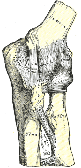
The ulnar collateral ligament of the elbow joint (UCL or internal lateral ligament) is a thick triangular band at the medial aspect of the elbow connecting the ulna to the humerus. It consists of two portions, an anterior and posterior, united by a thinner intermediate portion. Note that this ligament is also referred to as the medial collateral ligament and should not be confused with the lateral ulnar collateral ligament (LUCL).


The ulnar collateral ligament of the elbow joint is visible at center left.

Attachments and Course

The anterior portion, directed obliquely forward, is attached, above, by its apex, to the front part of the medial epicondyle of the humerus; and, below, by its broad base to the medial margin of the coronoid process of the ulna. The posterior portion, also of triangular form, is attached, above, by its apex, to the lower and back part of the medial epicondyle; below, to the medial margin of the olecranon. Between these two bands, a few intermediate fibers descend from the medial epicondyle to blend with a transverse band, which bridges across the notch between the olecranon and the coronoid process.

This ligament is in relation with the triceps brachii and flexor carpi ulnaris and the ulnar nerve, and gives origin to part of the flexor digitorum superficialis.

Attachments:


Ulnar_elbow.png (image/png)

Last Updated on September 27, 2026

🔗 Knowledge Graph & Clinical Correlates

OrthopaedicsOne Living Semantic Network

🛠️ Operative Techniques

  • Ulnohumeral Arthroplasty (Outerbridge-Kashiwagi) Shoulder and Elbow
  • Two Incision Technique for the Treatment of Distal Biceps Tendon Disruptions Shoulder and Elbow

🩺 Clinical Conditions & Indications

  • Valgus extension overload 1. High-Yield Summary Definition: Valgus extension overload (VEO) is an overuse impingement syndrome of the overhead athlete caused...
  • Ulnar nerve compression at the elbow 1. High-Yield Summary Cubital tunnel syndrome is the second most frequent compressive neuropathy of the upper extremity, trailing...
  • Ulnar collateral ligament injury of the elbow 1. High-Yield Summary Ulnar collateral ligament (UCL) injury of the elbow is a debilitating condition predominantly affecting overhead...
  • Triceps rupture 1. High-Yield Summary Rarity and Mechanism: Distal triceps tendon rupture is the least common of all major tendon...

🔬 Anatomy, Pathology & Diagnostics

  • Wrist joint Wrist and Carpus
  • Vertebra prominens Surface Anatomy Lumbar Spine
  • Vastus medialis Femoral Shaft
  • Vastus lateralis Femoral Shaft

🕸️ Semantic Relations (RDF)

ABOUT

  • Overview
  • FAQs
  • Managing Editors
  • Editorial Board
  • Content Partners
  • Participating Groups
  • Advisory Board
  • Sponsoring Organizations
  • Contact Us

ARTICLES

  • Arthroscopy
  • Foot and Ankle
  • Hand and Wrist
  • Hip and Knee
  • Infection
  • Joint Reconstruction
  • Musculoskeletal Medicine
  • Pediatrics
  • Shoulder and Elbow
  • Spine
  • Sports Medicine
  • Trauma
  • Tumor

REVIEWS

  • For Residents
  • For Medical Students

EBOOKS

  • PORT Notes
  • MSK Medicine
  • Tumor Library

COMMUNITY

  • Talks
  • Images
  • Viewpoints
  • Question Bank
  • Devices

PAGES

  • Residency
  • Fellowships
  • Associations
  • Companies
Copyright © 2012-2026 OrthopaedicsOne | Privacy Policy | Dr. Christian Veillette . Powered by: OrthoNet - Empowering the Future of Orthopaedic Care
Scroll to top Scroll to top Scroll to top