
Distal tibiofibular syndesmotic injuries occur in up to 20% of ankle fractures requiring internal fixation.1,2 Patients with ankle injuries with concomitant syndesmotic disruption have more pain and poorer function 1 year post-injury than those with ankle fractures without syndesmotic injury. This may reflect the severity of the initial injury; however, in their series of 25 ankles with postoperative X-ray and CT imaging, Gardner et al observed that up to 52% of syndesmoses were malreduced.
Failure to diagnose instability is one of the more common pitfalls in treating syndesmosis injuries. Diagnosis has classically been determined radiographically on AP and mortise projections. The standard criteria for a reduced syndesmosis measured 1cm proximal to the plafond on AP and mortise views are:
- Tibiofibular clear space of <6 mm
- Tibiofibular overlap of >6 mm or 42% of the width of the fibula on the AP view
- >1 mm of overlap on mortise view
When clinically suspicious, an external rotation stress view may demonstrate displacement compared to normal static radiographs. Intra-operatively, the direct lateral stress or “hook” test has been shown to be more reliable than the external rotation test.
Once diagnosed, another common pitfall is malreduction of the syndesmosis at the time of definitive fixation. Restoration of anatomic length and rotation of the fibula at the level of the syndesmosis are prerequisites to obtaining a satisfactory syndesmosis reduction. Clues to malreduction of the fibula and/or the syndesmosis include:
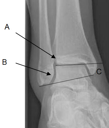
- Lateral shift of the talus with reference to the cortical density of the sulcus of the syndesmosis on a mortise view
- Non-congruence of the lateral talus relative to the distal fibula
- Abnormal talocrural angle (Figures 1a-1b)

Figure 1a. With anatomic reduction of the distal fibula, the lateral talus is in line with the sulcus of the syndesmosis (A), there is congruence of the distal fibula with the talus (B), and the talo-crural angle is 11 ± 4 º (C).
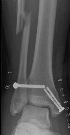
Figure 1b. With non-anatomic restoration of fibular length and rotation, syndesmosis malreduction is inevitable.
The syndesmosis can easily be assessed under direct visualisation from its anterior aspect to confirm the accuracy of reduction.
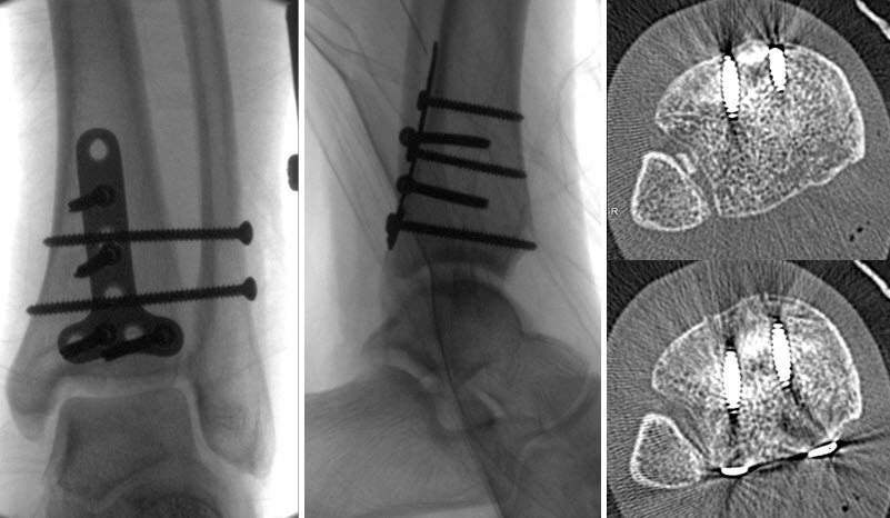
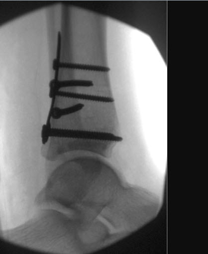
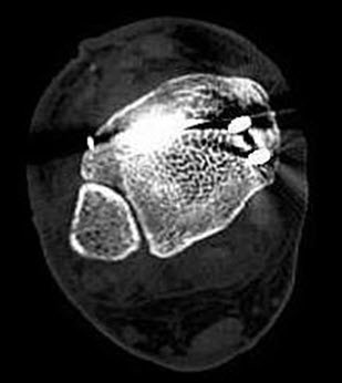
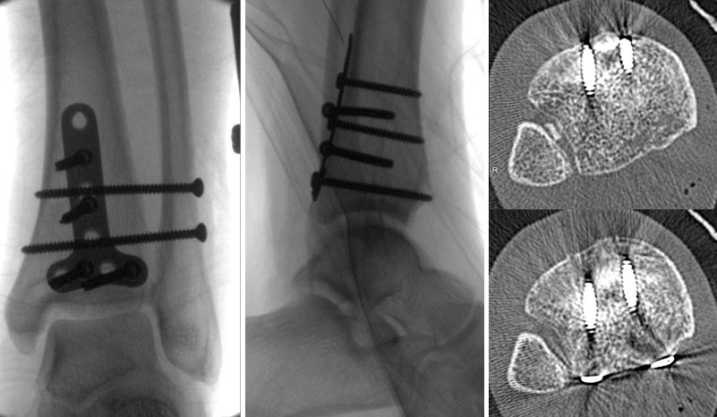
The lateral view is also important for confirming proper reduction of an unstable syndesmosis, as instability is greatest in the sagittal plane and thus better demonstrated on a lateral radiograph.7,8 The fibula is a posteriorly biased structure, but on a true lateral view, the fibula should not lie posterior to the tibia at the joint level. It is possible to satisfy the AP/mortise reduction criteria and yet have a malreduced syndesmosis (Figures 2a-b).
Figure 2a. After open reduction and internal fixation of a posterior malleolus and syndesmosis injury, the mortise appears reduced, but the fibula is posteriorly displaced on the lateral view. CT scan confirms syndesmosis malreduction with interposed loose body.
Figure 2b. Lateral view after removal of loose body and revision reduction of syndesmosis. Note that the posterior border of the fibula is anterior to the posterior border of the posterior malleolus.
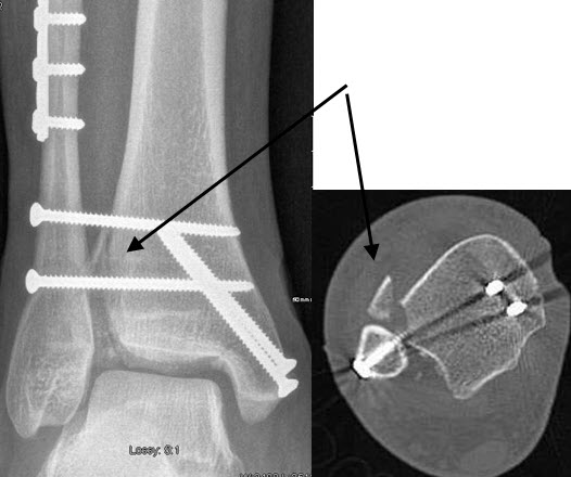
Fractures of the lateral aspect of the distal tibia, such as an anterolateral tibial fracture (Chaput fragment) can be potential pitfalls of the direct visualisation technique. If the syndesmosis reduction is based on visualisation of the anterior tibia and fibula, the syndesmosis will appear reduced when in fact it is anteriorly displaced (Figures 3a-b). If any doubt remains, a postoperative CT scan is indicated. Radiographs have been shown to have a sensitivity of 31% and a specificity of 83% for detection of syndesmosis malreduction when compared to CT scans based on AP and mortise radiographic criteria.
Figure 3a. Malreduced syndesmosis with an anterolateral tibial (Chaput) fracture.
Figure 3b. Syndesmosis reduced after reduction and fixation of the Chaput fragment.
The best method of fixation of the syndesmosis has not yet been determined. The current literature does not show a convincing advantage based on the type (rigid vs. flexible), size and number of screws, or the extent (three vs. four) of cortical fixation. Likewise, the evidence is inconclusive regarding the length of postoperative weight-bearing restriction or need for hardware removal. Two recent studies have shown benefit to removing intact screws;9,10 however, the removal of loose or broken screws is likely unnecessary. We prefer two 3.5-mm fully threaded screws across four cortices, 3 months non-weight-bearing, and removal of screws only if symptomatic.
Despite the frequency of these injuries and the wealth of related studies, the majority of controversies surrounding the fixation of syndesmosis injuries remain unresolved and require further study. Attention to avoid the common pitfalls of missed diagnosis and malreduction can predictably reduce the adverse effects on functional outcomes.
References
- Kennedy J.G., Johnson S.M., Collins S.L., et al. An evaluation of the Weber classification of ankle fractures. Injury 1998; 29: 577-80.
- Pettrone F.A., Gail M., Pee D., et al. Quantitative criteria for prediction of the results after displaced fracture of the ankle. J Bone Joint Surg (Am) 1983; 65-A: 667-77.
- Egol K.A., Pahk B., Walsh M., et al. Outcome after Unstable Ankle Fracture: Effect of Syndesmotic Stabilization. J Orthop Trauma 2010; 24: 7-11.
- Gardner M.J., Demetrakopoulos D., Briggs S.M., et al. Malreduction of the tibiofibular syndesmosis in ankle fractures. Foot Ankle Int 2006; 27: 788-792.
- Harper M.C., Keller T.S. A radiographic evaluation of the tibiofibular syndesmosis. Foot Ankle 1989; 10: 156-160.
- Stoffel K., Wysocki D., Baddour E., et al. Comparison of Two Intraoperative Assessment Methods for Injuries to the Ankle Syndesmosis. J Bone Joint Surg Am 2009; 91-A: 2646-52.
- Xenos J.S., Hopkinson W.J., Mulligan M.E., et al. The Tibiofibular Syndesmosis. Evaluation of the Ligamentous Structures, Methods of Fixation, and Radiographic Assessment. J Bone Joint Surg Am 1995; 77-A; 847-856.
- Candal-Couto J.J., Burrow D., Bromage S., et al. Instability of the tibio-fibular syndesmosis: have we been pulling in the wrong direction? Injury 2004; 35: 814-818.
- Miller A.N., Paul O., Boraiah S., et al. Functional Outcomes After Syndesmotic Screw Fixation and Removal. J Orthop Trauma 2010; 24: 12-16.
- Manjoo A., Sanders D., Tieszer C., et al. Functional and Radiographic Results of Patients with Syndesmotic Screw Fixation: Implications for Screw Removal. J Orthop Trauma 2010; 24: 2-6.
Reprinted with permission from the Spring 2010 issue of COA Bulletin
Attachments:

LiewFig1a.jpg (image/jpeg)
LiewFig1b.jpg (image/jpeg)
LiewFig2a.jpg (image/jpeg)
LiewFig2b.jpg (image/jpeg)
LiewFig3a.jpg (image/jpeg)
LiewFig3b.jpg (image/jpeg)
LiewFig2a.jpg (image/jpeg)
Last Updated on June 27, 2024
🔗 Knowledge Graph & Clinical Correlates
OrthopaedicsOne Living Semantic Network🛠️ Operative Techniques
- Wound coverage techniques Foot and Ankle
- Weil Lesser Metatarsal Shortening Osteotomy Foot and Ankle
- Varus and Valgus Lesser Toe Deformity Correction Foot and Ankle
- V-Y Advancement and FHL Transfer for the Chronic or Neglected Achilles Tendon Rupture – Raikin Foot and Ankle
🧭 Surgical Approaches & Danger Zones
- Transfibular Approach Ankle Arthrodesis Ankle Joint & Pilon
🩺 Clinical Conditions & Indications
- Torsional problems 1. High-Yield Summary Lower extremity torsional variations and rotational problems represent the most frequent reason for pediatric orthopaedic...
🔬 Anatomy, Pathology & Diagnostics
- Total Ankle Replacement using the Topez Total Ankle System Ankle Joint & Pilon
- Total ankle replacement (TAR) Ankle Joint & Pilon
- TNK Total Ankle Replacement Ankle Joint & Pilon