
Contents
- Name of bone
- Location/Articulation
- Muscle and ligament attachments
- Surface anatomy
- Radiography
- Physical examination
- Embryology
- Anomalies
- Injuries/Disorders
- Figures
Name of bone
Scapula
Location/Articulation
In anatomy, the scapula, omo, or shoulder blade is the bone that connects the humerus (arm bone) with the clavicle (collar bone). The scapula forms the posterior part of the shoulder girdle. In humans, it is a flat bone, roughly triangular in shape, placed on a posterolateral aspect of the thoracic cage.
Muscle and ligament attachments
The following muscles attach to the scapula:
Muscle | Direction | Region |
Pectoralis Minor | Insertion | Coracoid process |
Coracobrachialis | Origin | Coracoid process |
Serratus Anterior | Insertion | Medial border |
Triceps Brachii (long head) | Origin | Infraglenoid tubercle |
Biceps Brachii (short head) | Origin | Coracoid process |
Biceps Brachii (long head) | Origin | Supraglenoid tubercle |
Supscapularis | Origin | Subscapular fossa |
Rhomboid Major | Insertion | Medial border |
Rhomboid Minor | Insertion | Medial border |
Levator Scapulae | Insertion | Medial border |
Trapezius | Insertion | spine of scapula |
Deltoid | Origin | Spine of scapula |
Supraspinatus | Origin | Supraspinous fossa |
Infraspinatous | Origin | Infraspinous fossa |
Teres Minor | Origin | Lateral border |
Teres Major | Origin | Lateral border |
Lastissimus Dorsi (a few fibers) | Origin | Inferior angle |
Omohyoid | Origin | Superior border |
Surface anatomy
Costal (Front, Ventral, Anterior)
The costal, or ventral, surface presents a broad concavity, the subscapular fossa. The medial two-thirds of this fossa are marked by several oblique ridges, which run lateral and upward. The ridges give attachment to the tendinous insertions, and the surfaces between them to the shelby of the subscapularis. The lateral third of the fossa is smooth and covered by the fibers of this muscle.
At the upper part of the fossa is a transverse depression, where the bone appears to be bent on itself along a line at right angles to and passing through the center of the glenoid cavity, forming a considerable angle, called the subscapular angle. This gives greater strength to the body of the bone by its arched form, while the summit of the arch serves to support the spine and acromion
Dorsal (Back, Posterior)
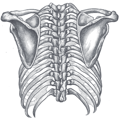
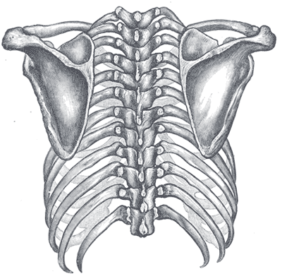
The dorsal surface is arched from above downward, and is subdivided into two unequal parts by the spine; the portion above the spine is called the supraspinatous fossa, and that below it is the infraspinous fossa
- The supraspinous fossa, the smaller of the two, is concave, smooth, and broader at its vertebral than at its humeral end. Its medial two thirds give origin to the supraspinatous
- The infraspinous fossa is much larger. Toward its vertebral margin, a shallow concavity is seen at its upper part; its center presents a prominent convexity, while near the axillary border is a deep groove that runs from the upper toward the lower part. The medial two thirds of the fossa give origin to the infraspinatous; the lateral third is covered by this muscle.
The dorsal surface is marked near the axillary border by an elevated ridge, which runs from the lower part of the glenoid cavity, downward and backward to the vertebral border, about 2.5 cm above the inferior angle. The ridge serves for the attachment of a fibrous septum, which separates the infraspinatous from the teres major and teres minor. The surface between the ridge and the axillary border is narrow in the upper two thirds of its extent, and is crossed near its center by a groove for the passage of the scapular circumflex vessels. It affords attachment to the teres minor
The lower third of the dorsal surfaces presents a broader, somewhat triangular surface, which gives origin to the teres major, and over which the latissimus dorsi glides. Frequently, the latter muscle takes origin by a few fibers from this part.
The broad and narrow portions alluded to above are separated by an oblique line, which runs from the axillary border, downward and backward, to meet the elevated ridge. To it is attached a fibrous septum that separates the teres muscles from each other.
Radiography
Enter radiographic images and landmarks
Physical examination
Enter relevant physical exam maneuvers for this bone
Embryology
Enter embryology for this bone
Anomalies
Enter anomalies for this bone
Injuries/Disorders
Because of its sturdy structure and protected location, scapular fractures are uncommon; when they do occur, they are an indication that severe chest trauma has occurred.
Figures

Figure 1. Right and left scapulae
Attachments:

Last Updated on June 27, 2024
🔗 Knowledge Graph & Clinical Correlates
OrthopaedicsOne Living Semantic Network🛠️ Operative Techniques
- Woodward repair of Sprengel Deformity Pediatrics
- Total scapular resections with endoprosthetic reconstruction Musculoskeletal Tumor
🩺 Clinical Conditions & Indications
- Traumatic brachical plexopathy 1. High-Yield Summary Traumatic brachial plexopathy represents a catastrophic peripheral nervous system injury predominantly caused by high-energy traction,...
🔬 Anatomy, Pathology & Diagnostics
- Wrist joint Wrist and Carpus
- Vertebra prominens Surface Anatomy Lumbar Spine
- Vastus medialis Femoral Shaft
- Vastus lateralis Femoral Shaft