
1. High-Yield Overview & Pathognomonic Essence
- Morphological Hallmark: An impaction fracture and wedge-shaped osteochondral depression located on the anteromedial aspect of the humeral head, situated at the articular margin immediately adjacent to the lesser tuberosity.
- Pathognomonic Significance: Represents the definitive osseous impaction lesion (McLaughlin defect) resulting from posterior glenohumeral dislocation, produced when the anteromedial humeral articular surface is violently driven against the dense posterior osseous glenoid rim. It is classically precipitated by violent, uncoordinated muscular contractions during epileptic seizures, high-voltage electrocution, or severe vehicular trauma involving adduction, flexion, and internal rotation forces.
- Critical Diagnostic & Surgical Implication: Notoriously overlooked on initial emergency evaluation, with missed diagnosis rates of 50% to 80% on solitary anteroposterior (AP) plain radiographs. Articular defect arc quantification via thin-slice computed tomography (CT) dictates surgical strategy according to the Hawkins classification: defects under 20% of the articular surface are stable following closed reduction and immobilization in external rotation; defects involving 20% to 40% require non-anatomical (modified McLaughlin lesser tuberosity transfer) or anatomical (osteochondral allograft) reconstruction to prevent recurrent engagement; and extensive defects exceeding 40% to 45% necessitate prosthetic arthroplasty.
2. Diagnostic Sign Summary Matrix
| Primary Modality | Anatomic Location | Primary Pathology | Pathognomonic Morphology | Diagnostic Utility & Sensitivity |
|---|---|---|---|---|
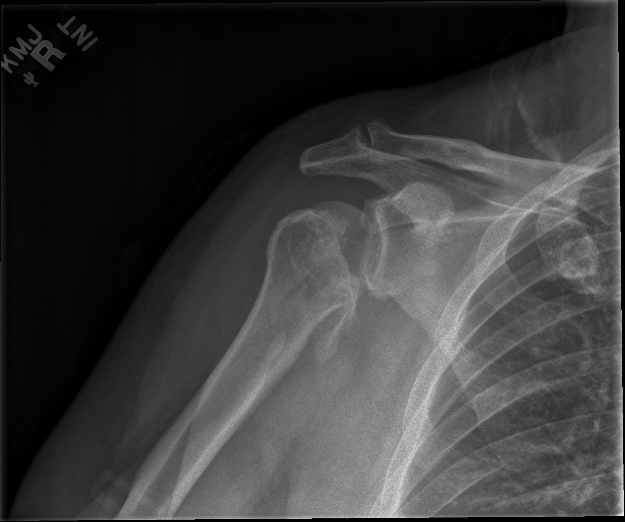
| Plain Radiography (AP, Axillary Lateral, Velpeau, Grashey) | Anteromedial Humeral Head & Glenohumeral Articulation | Locked Posterior Glenohumeral Dislocation | Vertical cortical condensation line (“trough line sign”), symmetric globular humeral head (“lightbulb sign”), absent elliptical glenohumeral overlap, and posterior humeral subluxation on axillary view | Solitary AP radiographs miss 50% to 80% of acute presentations; orthogonal axillary or Velpeau views elevate diagnostic sensitivity to >90%, confirming posterior displacement and anterior impaction |
| Computed Tomography (Thin-Slice Multiplanar CT & 3D Reformations) | Anteromedial Humeral Articular Surface & Posterior Glenoid Rim | Reverse Hill-Sachs Lesion (McLaughlin Defect) & Reverse Bankart Lesion | Sharply demarcated V-shaped or wedge-shaped anteromedial osteochondral compression defect, quantification of articular arc involvement, and associated posterior glenoid rim fracturing | Definitive gold standard (sensitivity and specificity approaching 100%); essential for measuring defect arc percentage, assessing engaging biomechanics, and planning reconstructive surgery |
| Magnetic Resonance Imaging (MRI & MR Arthrography) | Anterior Humeral Head, Posterior Labrocapsular Complex & Subscapularis Tendon | Posterior Shoulder Instability / Posterior Labrocapsular Disruption | Geographic subchondral bone marrow edema on T2/STIR sequences at the anteromedial head, osteochondral depression, posterior labral tear (reverse Bankart / Kim lesion), and subscapularis tendon continuity | Superior soft-tissue resolution (sensitivity 90-95% for chondrolabral pathology); differentiates acute marrow edema from chronic scarring and verifies rotator cuff and posterior capsule integrity |
3. Clinical Case Imaging & Standardized Figures

4. Pathophysiology & Biomechanical Mechanism
The reverse Hill-Sachs lesion, historically designated the McLaughlin lesion, represents a focal impaction fracture involving the anteromedial articular surface of the humeral head immediately adjacent to the lesser tuberosity. Harrison L. McLaughlin first described this structural depression in 1952 as the diagnostic counterpart to the classic anterior Hill-Sachs lesion, documenting that posterior displacement of the humeral head forces its anterior cartilaginous and subchondral surface violently against the dense, hard posterior osseous glenoid rim.
The biomechanical genesis of this injury is intimately linked to the muscular anatomy and force couples governing the shoulder girdle. The glenohumeral joint is uniquely susceptible to posterior dislocation during episodes of uncoordinated, massive muscular spasm, classically encountered during grand mal epileptic seizures, electroconvulsive therapy, or high-voltage electrical injuries (electrocution). Under these conditions, all muscle groups spanning the shoulder fire simultaneously. The internal rotators and adductors—predominantly the latissimus dorsi, pectoralis major, and subscapularis—possess significantly greater physiological cross-sectional area and mechanical advantage compared to the relatively weaker external rotators (infraspinatus and teres minor). The resulting net muscular force vector violently forces the humeral head into extreme internal rotation, adduction, and flexion.
As the humeral head is driven posteriorly past the posterior labrum and capsule, the dense cortical bone of the posterior glenoid rim acts as an anvil. The softer, cancellous subchondral bone of the anterior humeral head impacts directly against the posterior rim, creating a vertical, wedge-shaped impression defect. The depth and arc of this defect depend upon the magnitude of the impaction force, bone mineral density, and whether the dislocation remains acutely locked.
From a functional standpoint, the geometry of the reverse Hill-Sachs defect determines whether it behaves as an engaging or non-engaging lesion. An engaging defect aligns parallel to the posterior glenoid rim when the arm is brought into functional positions of internal rotation, forward flexion, and adduction. During these motions, the anterior edge of the defect slips over the posterior glenoid rim, dropping into the notch and leveraging the humeral head out of the glenoid cavity. This produces recurrent posterior instability or maintains a chronically locked posterior dislocation. Conversely, non-engaging defects remain clear of the posterior rim throughout physiological motion arcs and do not cause mechanical catching or recurrent subluxation.
5. Modality-Specific Imaging Findings
Plain Radiography
Conventional anteroposterior (AP) radiography of the shoulder is notoriously deceptive in posterior dislocations, frequently appearing deceptively normal or displaying subtle indirect signs that are easily misinterpreted by clinicians:
- The Trough Line Sign: Visualized on frontal AP projections as one or two dense vertical radiopaque lines traversing the anteromedial humeral head, oriented parallel to the medial humeral articular cortex. This vertical line represents the dense, compacted cortical margins of the impaction defect viewed en face on tangent to the radiographic beam.
- The Lightbulb Sign: Because the humeral head is locked in fixed internal rotation, the greater tuberosity is rotated anteriorly toward the midline. The humeral head loses its normal anatomic asymmetrical profile and projects symmetrically and spherically over the glenoid, resembling an incandescent electric lightbulb or an ice cream cone.
- Loss of Normal Glenohumeral Overlap: On a standard AP view, the normal elliptical overlapping shadow between the posterior glenoid rim and the medial humeral head (the half-moon overlap) is obliterated. The joint space may appear falsely widened (>6 mm, known as the positive “rim sign”) or exhibit abnormal medial displacement and overlap depending on the projection angle.
- The Vacant Glenoid Sign: The anterior half of the glenoid fossa appears empty and uncovered because the humeral head has migrated completely into the posterior extrascapular space.
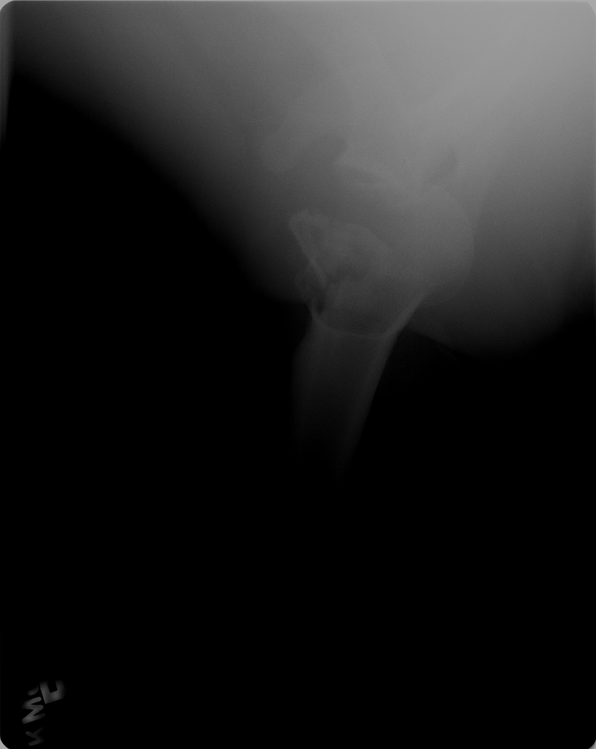
Because solitary AP radiographs result in high missed rates, mandatory orthogonal projections must be performed. The axillary lateral view is the single most critical projection, demonstrating unequivocal posterior displacement of the humeral head relative to the glenoid fossa and displaying the V-shaped anteromedial impaction defect locked onto the posterior glenoid rim. If acute pain and muscular spasm prevent arm abduction, a Velpeau axillary view (obtained with the patient leaning backward 30° over the film cassette with the arm resting in a sling) or a scapular Y view (showing the humeral head centered posterior to the intersection of the spine, acromion, and coracoid) provides definitive orthogonal confirmation.
Computed Tomography
Multi-detector computed tomography (CT) with thin-slice (1 mm or sub-millimeter) axial acquisitions and multiplanar coronal, sagittal, and 3D volume-rendered reformations represents the gold standard imaging modality. Axial CT slices clearly delineate the exact location, morphology, depth, and articular arc percentage of the reverse Hill-Sachs defect along the anteromedial humeral head. The defect appears as a sharp, wedge-shaped corticosubchondral impression defect with compressed trabecular bone adjacent to the bicipital groove and lesser tuberosity.
CT imaging allows precise quantification of articular surface involvement using the arc angle method. The angle subtended by the defect is measured relative to the total 180° circular arc of the normal humeral articular surface on the axial cut exhibiting maximum defect width. Furthermore, CT accurately evaluates concomitant osseous pathology, including posterior glenoid rim fracture (reverse bony Bankart lesion), posterior glenoid retroversion and dysplasia, and lesser tuberosity avulsion fractures. Post-processing 3D volume reconstructions with digital humeral head subtraction allow unobstructed en face evaluation of the glenoid articular surface, essential for comprehensive preoperative planning.
Magnetic Resonance Imaging & Ultrasound
Magnetic resonance imaging (MRI) and MR arthrography provide superior soft-tissue characterization while accurately assessing the osteochondral lesion. On fluid-sensitive sequences (T2-weighted fat-suppressed and STIR), an acute reverse Hill-Sachs lesion demonstrates prominent geographic subchondral bone marrow edema beneath the impacted articular surface, contrasting sharply with the low-signal cortical line. In chronic locked dislocations, marrow edema resolves and is replaced by low T1 and T2 signal corresponding to dense reparative subchondral sclerosis and fibrosis.
MRI is uniquely capable of assessing the integrity of the soft-tissue restraints, specifically the posterior labrocapsular complex and rotator cuff. Associated lesions frequently identified include posterior labral tears (reverse Bankart lesion), incomplete posterior labrocapsular avulsions with concealed retro-labral clefts (Kim lesions), tears of the posterior inferior glenohumeral ligament (PIGHL), and tears or avulsions of the subscapularis tendon from the lesser tuberosity. High-resolution diagnostic ultrasound has limited utility in quantifying deep osseous defects due to acoustic shadow artifact from cortical bone, but dynamic posterior sonography can demonstrate posterior joint subluxation during gentle external rotation maneuvers and can verify subscapularis tendon integrity.
6. Diagnostic Utility, Sensitivity & Specificity
The diagnostic utility of imaging in posterior shoulder dislocation and reverse Hill-Sachs lesions depends fundamentally on obtaining appropriate orthogonal views. Conventional AP plain radiographs exhibit a notorious failure rate: published retrospective series demonstrate that between 50% and 80% of posterior dislocations are missed at initial emergency department presentation when evaluated solely with frontal projections. The subtle nature of the lightbulb and trough line signs often leads clinicians to misdiagnose the condition as frozen shoulder, rotator cuff contusion, or post-ictal paresis.
When orthogonal projections—specifically the axillary lateral or Velpeau view—are included in the radiographic trauma series, the sensitivity of plain radiography for identifying posterior dislocation and the associated anteromedial humeral defect rises to over 90% to 95%. Thin-slice multiplanar CT achieves sensitivity and specificity approaching 100%, serving as the definitive reference standard. CT provides flawless identification of the reverse Hill-Sachs defect, accurate calculation of the articular arc involvement percentage, and precise characterization of posterior glenoid rim integrity. The positive predictive value of a concurrent trough line sign and lightbulb sign on plain radiography is high (exceeding 95%), but the absence of these signs on an isolated AP radiograph can never be used to exclude posterior dislocation.
7. Differential Diagnosis & Radiographic Mimics
| Sign / Condition | Distinguishing Radiographic Features | Clinical Presentation | Key Differentiating Test |
|---|---|---|---|
| Reverse Hill-Sachs Lesion (McLaughlin Defect) | Anteromedial humeral head impaction defect at/adjacent to lesser tuberosity; associated with posterior dislocation, trough line sign, and lightbulb sign on AP | Seizure, electrocution, or adduction trauma; arm locked in rigid internal rotation; complete inability to passively externally rotate past neutral | Axillary lateral radiograph and axial CT confirming posterior glenohumeral dislocation and anteromedial osteochondral notch |
| Classic Hill-Sachs Lesion | Posterolateral superior humeral head impaction defect; associated with anterior glenohumeral dislocation and anterior labral/Bankart lesions | Abduction, external rotation, and extension trauma; arm held in slight abduction and external rotation; anterior fullness on palpation | Axillary radiograph and axial CT demonstrating anterior humeral displacement and posterolateral humeral head notch |
| Anatomical Humeral Head Sulcus (Normal Bare Area) | Smooth, shallow physiological groove at the junction of the articular cartilage and posterior humeral neck, 1.5 to 2.0 cm below superior margin; lacks cortical impaction or step-off | Asymptomatic, incidental anatomical finding on cross-sectional imaging; full, painless shoulder motion with normal stability | MRI or CT showing smooth cortical contour without subchondral trabecular impaction, bone marrow edema, or joint subluxation |
| Fracture of the Anatomical or Surgical Neck | Discontinuous fracture line traversing the anatomical or surgical neck; may feature cortical displacement or comminution, but humeral head articular dome remains spherical without focal wedge notch | Elderly fall or direct shoulder trauma; extensive ecchymosis of chest wall and arm; marked pain without locked internal rotation | Multiplanar CT demonstrating cortical fracture lines across the humeral neck with preserved spherical articular contour |
| Avascular Necrosis (AVN) of Humeral Head / Subchondral Collapse | Subchondral radiolucent crescent sign, patchy sclerosis, and articular collapse confined to the superior weight-bearing dome of the humeral head; normal glenohumeral alignment | Insidious, progressive shoulder pain; history of corticosteroid therapy, sickle cell disease, alcohol abuse, or previous humeral fracture | MRI demonstrating serpentine demarcation line, subchondral collapse at superior apex, and absence of anteromedial wedge defect or dislocation |
8. Clinical Significance & Management Implications
The detection and dimensional quantification of a reverse Hill-Sachs lesion represents the single most critical factor guiding clinical decision-making and surgical reconstruction in posterior shoulder instability. Treatment algorithms are dictated primarily by the size of the articular defect (expressed as a percentage of the total humeral articular arc), the duration of locked dislocation, the age and functional demands of the patient, and the structural integrity of the posterior glenoid:
- Small Defects (<20% of Articular Arc): Acute posterior dislocations associated with small anteromedial defects involving less than 20% of the articular surface are biomechanically stable following closed reduction under adequate sedation or general anesthesia. Once reduced, the arm is immobilized in neutral to 15-20° of external rotation and slight extension using a dedicated external rotation brace for 3 to 4 weeks. Immobilization in internal rotation is strictly avoided, as it places the anteromedial defect directly against the posterior glenoid rim and predisposes to recurrent posterior redislocation.
- Medium Defects (20% to 40% of Articular Arc) — Non-Anatomical Reconstruction: Defects occupying between 20% and 40% of the humeral articular surface are prone to recurrent posterior instability and engagement during activities of daily living. The classic McLaughlin procedure involves open detachment of the subscapularis tendon insertion from the lesser tuberosity and direct transosseous suturing of the tendon into the debrided anteromedial defect, non-anatomically filling the void and preventing engagement. The modified McLaughlin (Neer) procedure mobilizes the lesser tuberosity via an osteotomy with the attached subscapularis tendon and transposes the bone block into the reverse Hill-Sachs defect, securing it with bicortical cancellous lag screws. This provides robust bone-to-bone healing, preserves native subscapularis tendon length, and demonstrates superior long-term union rates compared to soft-tissue transfer alone.
- Medium Defects (20% to 40% of Articular Arc) — Anatomical Reconstruction: In young, active patients desiring joint-preserving anatomical reconstruction, segmental fresh-frozen osteochondral allografting (pioneered by Gerber and Lambert) restores native articular sphericity and cartilage congruity. The graft (typically sculpted from a donor femoral head or humeral head) is press-fit and rigidly fixed with headless compression screws. Alternatively, for acute, uncomminuted impaction defects identified within two to three weeks of injury, disimpaction and humeroplasty (percutaneous or open elevation of the depressed osteochondral fragment combined with structural subchondral bone graft packing) can restore native articular contours.
- Large Defects (>40% to 45% of Articular Arc) — Prosthetic Arthroplasty: Extensive defects involving greater than 40% to 45% of the humeral head articular surface compromise native joint reconstructibility, carrying an unacceptable risk of post-traumatic avascular necrosis, progressive cartilage degeneration, and construct failure if joint preservation is attempted. In physiologically active patients with preserved, healthy glenoid cartilage and an intact posterior glenoid rim, prosthetic hemiarthroplasty restores joint stability, with humeral component retroversion carefully adjusted to neutral (0° to 10° of retroversion rather than standard 30°) to counteract posterior subluxation. In elderly patients, chronic locked dislocations exceeding 6 months duration, or cases complicated by severe posterior glenoid bone loss and cartilage erosion, reverse total shoulder arthroplasty (rTSA) provides reliable pain relief and eliminates the risk of posterior prosthesis dislocation by establishing a semi-constrained, biomechanically stable inverted articulation.
- The Chronicity Factor & Danger of Iatrogenic Fracture: Dislocations chronically locked for more than 3 to 6 weeks undergo severe capsular contraction, soft-tissue fibrosis, and disuse osteopenia. Closed reduction in chronic locked dislocations is strictly contraindicated due to the extreme risk of iatrogenic spiral humeral fracture; open surgical reduction via an extended deltopectoral approach is mandatory.
9. Master Class Pro-Tip
Master Class Pro-Tip: In any patient presenting with severe shoulder pain and an absolute mechanical block to passive external rotation following an epileptic seizure, high-voltage electrical shock, or high-energy trauma, assume a locked posterior glenohumeral dislocation with an engaging reverse Hill-Sachs lesion until proven otherwise. Because standard anteroposterior radiographs fail to detect posterior dislocation in over 50% of cases, an orthogonal axillary lateral or Velpeau view is mandatory, and thin-slice multiplanar CT should be obtained immediately to quantify the anteromedial defect arc. If the dislocation has been locked for more than two to three weeks, never attempt forceful closed reduction or rotational manipulation, as the engaged humeral head acts as a rigid fulcrum against the posterior glenoid rim, producing catastrophic spiral fractures of the proximal humeral shaft; open reduction and anatomical or non-anatomical defect filling is required.
Master Class Radiological Pearl: Reverse Hill-Sachs Lesion and Posterior Shoulder Dislocation
10. Evidence-Based Synthesis
| Author & Year | Study Design | Sample Size | Key Findings & Diagnostic Accuracy | Clinical Implication | Level of Evidence | DOI |
|---|---|---|---|---|---|---|
| McLaughlin HL (1952) | Seminal Surgical Technique & Case Series | N = 22 locked posterior shoulder dislocations | First systematically described the anteromedial humeral head impaction defect (‘McLaughlin lesion’) created by impaction against the posterior glenoid rim in posterior shoulder dislocation; pioneered open reduction and stabilization via subscapularis tendon transfer into the defect to prevent redislocation. | Established anterior humeral head impression defect filling as the standard biomechanical principle to neutralize engaging reverse Hill-Sachs defects. | Level V | DOI |
| Hawkins RJ et al. (1987) | Seminal Case Series & Surgical Classification | N = 41 locked posterior shoulder dislocations | Established the foundational defect-size-based treatment algorithm (Hawkins classification): defects <20% treated with closed reduction and external rotation immobilization; defects 20-40% treated with lesser tuberosity transfer (modified McLaughlin / Neer procedure); defects >40-45% managed with prosthetic arthroplasty. | Created the universally accepted clinical framework correlating articular impression defect percentage with surgical selection and joint reconstructibility. | Level IV | DOI |
| Gerber C & Lambert SM (1996) | Landmark Prospective Case Series | N = 4 shoulders (segmental humeral head defects 30-50%) | Pioneered anatomical structural reconstruction of large reverse Hill-Sachs defects (30-50% articular arc) using fresh-frozen femoral head osteochondral allografts contoured to the defect and fixed with cancellous lag screws, successfully restoring humeral head sphericity and joint stability. | Provided an anatomical joint-preserving alternative to prosthetic hemiarthroplasty in young, high-demand patients with substantial articular cartilage destruction. | Level IV | DOI |
| Longo UG et al. (2020) | Systematic Review & Evidence Synthesis | Systematic review of 33 studies (N = 1,027 patients) | Synthesized outcomes across operative modalities for posterior shoulder instability and locked dislocations; confirmed that addressing anterior humeral bone defects (reverse Hill-Sachs) and posterior glenoid bone loss is critical to preventing recurrent posterior instability and catastrophic failure. | Highlights that unaddressed reverse Hill-Sachs defects >=20% result in unacceptable rates of recurrent posterior subluxation, mandating structural or non-anatomical defect filling. | Level I | DOI |
| Prigmore B et al. (2024) | Systematic Review & Meta-Analysis | Systematic review of 22 studies (N = 258 shoulders) | Evaluated osteochondral allograft transplantation for focal humeral articular defects, demonstrating high graft incorporation (88%), significant pain relief, and restoration of glenohumeral stability in patients with reverse Hill-Sachs defects involving 20% to 45% of the humeral head. | Validates osteochondral allografting as an effective, durable joint-preserving reconstructive option that delays or eliminates the need for early prosthetic arthroplasty in active patients. | Level I | DOI |
| Al-Mohrej OA et al. (2026) | Systematic Review & Clinical Decision Algorithm | Comprehensive systematic literature analysis | Formulated an evidence-based clinical decision algorithm integrating chronological duration of dislocation, patient physiologic age, glenoid articular integrity, and exact reverse Hill-Sachs defect sizing on multiplanar CT. | Recommends joint-preserving tuberosity transfers or allografts for defects 20-40% under 6 months duration, while prioritizing reverse or anatomic arthroplasty for defects >40% or chronic dislocations with degenerative glenoid wear. | Level IV | DOI |
| Duan D et al. (2026) | Clinical Case Series & Systematic Synthesis | N = 18 patients with locked posterior dislocation | Demonstrated that the modified McLaughlin procedure (lesser tuberosity osteotomy and screw fixation into the reverse Hill-Sachs defect) achieved 100% stable union, eliminated recurrent instability, and restored functional external rotation in defects comprising 20% to 45% of the humeral head. | Confirms bone-to-bone healing in the modified McLaughlin technique provides superior long-term fixation stability compared to soft-tissue subscapularis tenodesis alone. | Level IV | DOI |
| De Giorgi S et al. (2026) | Multicenter Cohort & Reconstructibility Framework | N = 42 locked posterior dislocations | Proposed a structured reconstructibility score assessing humeral head cartilage viability, reverse Hill-Sachs defect depth and arc, glenoid erosion, and dislocation chronicity (>6 weeks vs <6 weeks) to guide decision between joint preservation and arthroplasty. | Provides objective radiographic and surgical benchmarks to avoid high failure rates associated with attempting joint preservation in non-reconstructible, chronically locked dislocations. | Level IV | DOI |
| Wang X et al. (2026) | Retrospective Comparative Case Series | N = 15 complex fracture-dislocations | Evaluated outcomes of combining lesser tuberosity transfer with proximal humeral locking plate fixation for fracture-dislocations featuring substantial reverse Hill-Sachs impaction defects; reported excellent osseous union, stability, and Constant scores at mean 24-month follow-up. | Establishes the safety and biomechanical efficacy of combining tuberosity transposition with rigid internal fixation in complex fracture-dislocation patterns. | Level IV | DOI |
| Moroder P et al. (2026) | Matched Comparative Imaging Cohort Study | N = 80 matched shoulders (posterior vs anterior instability) | Matched CT/MRI morphometric analysis demonstrated that shoulders with posterior instability and reverse Hill-Sachs lesions exhibit significantly greater glenoid retroversion (mean -9.2° vs -2.8°) and a flatter acromial slope, altering joint kinematics and predisposing to posterior impaction. | Emphasizes that preoperative cross-sectional imaging must quantify underlying bony glenoid retroversion and morphology in addition to the humeral impaction defect. | Level III | DOI |
11. References
- McLaughlin HL. Posterior dislocation of the shoulder. J Bone Joint Surg Am. 1952;34-A(3):584-590. DOI [Level V: Seminal Surgical Technique & Landmark Case Series]
- Hawkins RJ, Neer CS 2nd, Pianta RM, Mendoza FX. Locked posterior dislocation of the shoulder. J Bone Joint Surg Am. 1987;69(1):9-18. DOI [Level IV: Seminal Case Series & Classification]
- Gerber C, Lambert SM. Allograft reconstruction of segmental defects of the humeral head for the treatment of chronic locked posterior dislocation of the shoulder. J Bone Joint Surg Am. 1996;78(3):376-382. DOI [Level IV: Landmark Case Series]
- Longo UG, Ciuffreda M, Locher J, et al. Posterior shoulder instability: a systematic review. Br Med Bull. 2020;134(1):34-53. DOI [Level I: Systematic Review / Meta-Analysis]
- Prigmore B, Tabbaa S, Crawford DC. Osteochondral Allograft Transplantation in the Shoulder: A Systematic Review of Indications and Outcomes. Cartilage. 2024;15(1):3-15. DOI [Level I: Systematic Review / Meta-Analysis]
- Al-Mohrej OA, Al-Kenani NS, Al-Hussainan TS, et al. Chronic posterior shoulder dislocation: Current evidence, treatment options, and a decision-making algorithm. J Orthop. 2026;55:102-110. DOI [Level IV: Systematic Review & Clinical Decision Algorithm]
- Duan D, Zhang L, Li M, et al. Surgical treatment of locked posterior shoulder dislocations with modified McLaughlin procedure: Case series and review of treatment possibilities of reverse Hill-Sachs defect. Chin J Traumatol. 2026;29(4):231-238. DOI [Level IV: Case Series & Systematic Literature Synthesis]
- De Giorgi S, Carrozzo M, Notarnicola A, et al. From reduction to replacement: a reconstructibility-based framework for chronic locked shoulder dislocations. Int Orthop. 2026;50(10):2541-2550. DOI [Level IV: Multicenter Cohort & Surgical Framework]
- Wang X, Chen Y, Liu Z, et al. Modified McLaughlin procedure combined with proximal humeral locking plate for proximal humeral fractures with posterior shoulder dislocation and reverse Hill-Sachs lesion: a retrospective case series. Front Surg. 2026;13:1881342. DOI [Level IV: Retrospective Case Series]
- Moroder P, Ernstbrunner L, Akgun D, et al. Posterior shoulder instability shows a higher, flatter acromion, and increased glenoid retroversion compared with anterior dislocation: A matched computed tomography/magnetic resonance imaging analysis. J ISAKOS. 2026;11(2):125-132. DOI [Level III: Comparative Imaging Cohort Study]
Last Updated on September 28, 2026
🔗 Knowledge Graph & Clinical Correlates
OrthopaedicsOne Living Semantic Network🛠️ Operative Techniques
- Turf Toe Injuries Sports Medicine
- Total Shoulder Arthroplasty for Glenohumeral Arthritis with an Intact Rotator Cuff Shoulder and Elbow
- The Role of Tendon Transfers in Massive Rotator Cuff Tears Shoulder and Elbow
- The Disabled Throwing Shoulder Sports Medicine
🩺 Clinical Conditions & Indications
- Turf toe Introduction Despite its rather benign namesake, turf toe is a incapacitating injury that plagues many athletes who participate...
- Therapeutic exercise for the shoulder Therapeutic exercise for the shoulder Range of Motion and Flexibility Pendulums Table Slides into flexion, external rotation, and...
- The cross-sectional study 1. High-Yield Summary Fundamental Architecture: A cross-sectional study (prevalence study or transverse survey) is an observational epidemiological design...
- Subacromial impingement 1. High-Yield Summary Subacromial impingement, now increasingly designated subacromial pain syndrome (SAPS) or rotator cuff-related shoulder pain (RCRSP),...
🔬 Anatomy, Pathology & Diagnostics
- Trough Line Glenohumeral Joint & Rotator Cuff
- The Role of Periosteum in Cartilage Repair General Musculoskeletal
- The morphology of the acromion according to Bigliani Glenohumeral Joint & Rotator Cuff
- The Biomechanical Case for Labral Débridement General Musculoskeletal