• O1 Network
    • Articles
    • For Residents
    • For Medical Students
    • PORT Notes
    • MSK Medicine
    • Tumor Library
    • Cases
    • Talks
    • Images
    • Viewpoints
    • Question Bank
    • Devices
    • Residency
    • Fellowships
    • Associations
    • Companies
ORTHOPAEDICSONE ARTICLES
OrthopaedicsOne Articles
  • Articles
    • Specialty
      • Arthroscopy
      • Foot and Ankle
      • Hand and Wrist
      • Hip and Knee
      • Infection
      • Joint Reconstruction
      • Musculoskeletal Medicine
      • Pediatrics
      • Shoulder and Elbow
      • Spine
      • Sports Medicine
      • Trauma
      • Tumor
    • Regional
      • Anatomy
      • Basic Science
      • Classifications
      • Diagnostic Tests
      • Evidence-Based Orthopaedics
      • Operative Techniques
      • Pain Management
      • Physical Examination
      • Pharmacology
      • Radiology
      • Rehabilitation
      • Rheumatology
      • Surgical Approaches
    • Professional
      • Bioethics
      • Economics and Health Policy
      • Epidemiology and Biostatistics
      • History of Orthopaedics
      • Informatics
      • Medicolegal
      • Mobile and Wireless
      • Practice Management
  • Reviews
    • For Residents
    • For Medical Students
  • eBooks
    • PORT Notes
    • MSK Medicine
    • Tumor Library
  • Community
    • Cases
    • Talks
    • Images
    • Viewpoints
    • Question Bank
    • Devices
  • Pages
    • Residency
    • Fellowships
    • Associations
    • Companies
  • About O1
    • Overview
    • FAQs
    • Managing Editors
    • Editorial Board
    • Content Partners
    • Participating Groups
    • Advisory Board
    • Sponsoring Organizations
    • Contact Us
  • Click to open the search input field Click to open the search input field Search
  • Menu Menu

Pronator teres

Origin

Humeral head: medial epicondyle of humerus (common flexor tendon)

Ulnar head: coronoid process of ulna

Insertion

Radius

Action(s)

Pronation of forearm, flexes elbow

Nerve Supply

Median nerve

Arterial Supply

Ulnar artery and radial artery

Physical Exam

Enter physical examination maneuvers for muscle

Clinical Importance

“Pronator teres syndrome” is one common cause of wrist pain.

Disease States

Enter links to pages where muscle involved

Discussion

Structure:

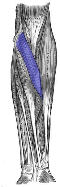
The pronator teres has two heads–humeral and ulnar.

The humeral head, the larger and more superficial, arises immediately above the medial epicondyle of the humerus and from the common flexor tendon.

The ulnar head is a thin fasciculus, which arises from the medial side of the coronoid process of the ulna, and joins the preceding at an acute angle.

The median nerve enters the forearm between the two heads of the muscle, and is separated from the ulnar artery by the ulnar head.

The muscle passes obliquely across the forearm, and ends in a flat tendon, which is inserted into a rough impression at the middle of the lateral surface of the body of the radius, just below the insertion of the supinator.

The lateral border of the muscle forms the medial boundary of triangular hollow known as the cubital fossa, which is situated in front of the elbow-joint.

Variations:

Occasionally, the ulnar head is absent. Also, additional slips from the medial intermuscular septum, from the biceps brachii, and from the brachialis occasionally occur.

Figures

Click thumbnail for larger image

Credits:

From Wikipedia:
Pronator teres

Attachments:


Pronator teres.png (image/png)

Last Updated on June 27, 2024

🔗 Knowledge Graph & Clinical Correlates

OrthopaedicsOne Living Semantic Network

🛠️ Operative Techniques

  • Ulnohumeral Arthroplasty (Outerbridge-Kashiwagi) Shoulder and Elbow
  • Two Incision Technique for the Treatment of Distal Biceps Tendon Disruptions Shoulder and Elbow

🩺 Clinical Conditions & Indications

  • Valgus extension overload 1. High-Yield Summary Definition: Valgus extension overload (VEO) is an overuse impingement syndrome of the overhead athlete caused...
  • Ulnar nerve compression at the elbow 1. High-Yield Summary Cubital tunnel syndrome is the second most frequent compressive neuropathy of the upper extremity, trailing...
  • Ulnar collateral ligament injury of the elbow 1. High-Yield Summary Ulnar collateral ligament (UCL) injury of the elbow is a debilitating condition predominantly affecting overhead...
  • Triceps rupture 1. High-Yield Summary Rarity and Mechanism: Distal triceps tendon rupture is the least common of all major tendon...

🔬 Anatomy, Pathology & Diagnostics

  • Wrist joint Wrist and Carpus
  • Vertebra prominens Surface Anatomy Lumbar Spine
  • Vastus medialis Femoral Shaft
  • Vastus lateralis Femoral Shaft

🕸️ Semantic Relations (RDF)

ABOUT

  • Overview
  • FAQs
  • Managing Editors
  • Editorial Board
  • Content Partners
  • Participating Groups
  • Advisory Board
  • Sponsoring Organizations
  • Contact Us

ARTICLES

  • Arthroscopy
  • Foot and Ankle
  • Hand and Wrist
  • Hip and Knee
  • Infection
  • Joint Reconstruction
  • Musculoskeletal Medicine
  • Pediatrics
  • Shoulder and Elbow
  • Spine
  • Sports Medicine
  • Trauma
  • Tumor

REVIEWS

  • For Residents
  • For Medical Students

EBOOKS

  • PORT Notes
  • MSK Medicine
  • Tumor Library

COMMUNITY

  • Talks
  • Images
  • Viewpoints
  • Question Bank
  • Devices

PAGES

  • Residency
  • Fellowships
  • Associations
  • Companies
Copyright © 2012-2026 OrthopaedicsOne | Privacy Policy | Dr. Christian Veillette . Powered by: OrthoNet - Empowering the Future of Orthopaedic Care
Scroll to top Scroll to top Scroll to top