
1. High-Yield Overview & Pathognomonic Essence
- Morphological Hallmark: The pneumoarthrogram sign (joint vacuum phenomenon) manifests as a sharply defined, curvilinear, crescentic radiolucency containing physiologic gas (predominantly molecular nitrogen) that conforms to and outlines the articular cartilage contour of a synovial joint, most classically observed in the hip and glenohumeral joints during mechanical traction or positional distraction.
- Pathognomonic Significance: The sign pathognomonically denotes the complete absence of a significant joint effusion (a dry, non-effused joint space). In the presence of joint distraction, the sudden expansion of a closed synovial cavity creates subatmospheric intra-articular pressure, provoking nitrogen outgassing from synovial fluid under Henry’s law.
- Critical Diagnostic Utility: Elicitation of the pneumoarthrogram sign provides a near-100% negative predictive value for joint effusion, serving as an invaluable non-invasive clinical discriminator to exclude acute septic arthritis or toxic synovitis in the pediatric irritable hip, while also providing fluoroscopic confirmation of successful articular separation during hip arthroscopy central compartment entry.
2. Diagnostic Sign Summary Matrix
| Primary Modality | Anatomic Location | Primary Pathology | Pathognomonic Morphology | Diagnostic Utility & Sensitivity |
|---|---|---|---|---|
| Plain Radiography (AP with Axial Traction) | Pediatric or Adult Hip Joint | Absence of joint effusion / physiologic vacuum cavitation | Curvilinear, crescentic gas-density lucency outlining the superolateral femoral head articular cartilage | Highly specific (>98%) for excluding joint effusion; traction-assisted sensitivity reaches 70% to 85% in non-effused joints |
| Plain Radiography (AP in External Rotation) | Glenohumeral Joint | Absence of joint effusion / physiologic vacuum cavitation under capsular tension | Sharply demarcated radiolucent gas crescent between the humeral head articular surface and the glenoid labrum | Distinguishes physiologic vacuum from rotator cuff arthropathy or effusive capsulitis; high diagnostic specificity |
| Dynamic Traction Fluoroscopy | Central Compartment of Hip (Arthroscopic positioning) | Articular coaptation seal breakdown under axial distraction (15-20 kg / 400-600 N) | Sudden appearance of a radiolucent vacuum cleft (2.5-4.2 mm distraction gap) prior to fluid infusion | Real-time operative verification of adequate distraction, preventing iatrogenic chondral scuffing during central portal access |
| Computed Tomography (CT) | Peripheral synovial joints (Hip, Shoulder, Knee, Sternoclavicular) | Micro-cavitation and gas release in non-effused joint under positioning stress | True gas attenuation (-900 to -1000 Hounsfield Units) conforming precisely to intact chondral surfaces without periarticular gas or soft-tissue stranding | 100% sensitivity for gas detection; crucial for ruling out open capsular disruption, necrotizing infection, or gas gangrene |
| Magnetic Resonance Imaging (MRI) | Glenohumeral joint (External rotation), Hip joint (FAI / Frog-leg positioning) | Localized distraction or levering of femoral/humeral head against the acetabular or glenoid rim | Complete signal void on all pulse sequences (T1, T2, PD) with prominent susceptibility “blooming” artifact on gradient-recalled-echo (GRE) sequences | High specificity; prevents misinterpretation as intra-articular loose bodies, calcific tendinopathy, or chondrocalcinosis |
3. Clinical Case Imaging & Standardized Figures

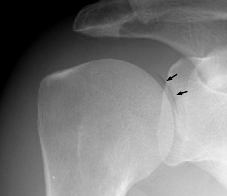
4. Pathophysiology & Biomechanical Mechanism
The pneumoarthrogram sign represents a direct physical manifestation of negative intra-articular hydrostatic pressure and gas cavitation in an enclosed, non-effused synovial joint. Under baseline physiological resting conditions, the joint capsule constitutes a hermetically sealed, non-compliant chamber. The healthy joint contains a minimal volume of synovial fluid (typically 0.5 to 1.5 mL in the hip or shoulder), which serves as a viscous lubricant and maintains a slightly subatmospheric baseline resting pressure of -2 to -5 cmH2O (-1.5 to -3.8 mmHg). This negative resting pressure contributes significantly to passive articular stability by establishing an atmospheric suction seal between congruent cartilage surfaces.
Synovial fluid exists in passive chemical equilibrium with the surrounding periarticular microvasculature and interstitial fluid, containing dissolved atmospheric gases: predominantly molecular nitrogen (N2, 78% to 80%), oxygen (O2, 15% to 18%), and carbon dioxide (CO2, 2% to 5%). According to Henry’s law of gas solubility, the concentration of a gas dissolved in a biological liquid is directly proportional to the partial pressure of that gas above or within the solution (C = k · P).
When external longitudinal traction or positional distraction is applied across a joint (such as 15 to 20 kg of manual axial leg traction or glenohumeral external rotation), the rigid osseous articular surfaces are forced apart while the tough fibrous capsule remains intact. Because synovial fluid volume is minimal and virtually incompressible, this sudden volumetric expansion causes intra-articular hydrostatic pressure to plunge precipitously into deeply negative subatmospheric territory, often dropping below -100 to -200 mmHg (-130 to -270 cmH2O). When the local pressure falls below the vapor and dissociation threshold of the dissolved gases, the liquid phase becomes abruptly supersaturated, driving instantaneous gas cavitation (effervescence) out of solution into the distracted articular cleft.
Molecular nitrogen constitutes over 90% to 92% of the liberated gas pocket. Because nitrogen possesses a low biological solubility coefficient and diffuses across synovial capillary membranes much more slowly than carbon dioxide or oxygen, the nitrogen gas pocket persists as a well-defined radiographic lucency throughout the duration of applied distraction. Upon releasing traction, the articular surfaces recoapt, intra-articular pressure normalizes, and the nitrogen gas is gradually redissolved back into the synovial liquid and capillary circulation.
Crucially, the presence of a joint effusion completely abolishes the vacuum phenomenon. In an effused, inflamed, or purulent joint (such as septic arthritis, toxic synovitis, or acute hemarthrosis), the intra-articular cavity contains an abnormal volume of fluid (often 10 to 50 mL or more). When axial traction is applied to an effused joint, this fluid freely shifts and redistributes across the joint recesses to occupy the distraction gap, preventing the generation of extreme negative pressures. Furthermore, pathological effusions elevate baseline intra-articular resting pressure to high positive levels (+20 to +60 cmH2O or greater). External traction forces cannot depress this elevated pressure below the critical subatmospheric threshold required for nitrogen cavitation. Consequently, nitrogen outgassing cannot occur in the presence of an effusion, establishing the pneumoarthrogram sign as a highly reliable marker of a dry, non-effused joint cavity.
5. Modality-Specific Imaging Findings
Plain Radiography
On standard anteroposterior (AP) radiographs of the hip or shoulder, the pneumoarthrogram sign appears as a thin, crisp, curvilinear crescent of air-density radiolucency hugging the articular surface. In the hip, the lucency is most commonly visualized along the superolateral margin of the femoral head cartilage under manual axial traction (10 to 15 kg in pediatric patients, 15 to 20 kg in adults) or on frog-leg lateral projections where the femoral neck levers against the acetabular rim. In the glenohumeral joint, external rotation positioning tightens the anterior capsule and distracts the posterior articular surface, rendering the crescent visible between the humeral head and glenoid fossa. The sign is strictly dynamic and transient; releasing traction allows the opposing cartilage surfaces to recoapt, instantly extinguishing the gas shadow.
Computed Tomography (CT)
Computed tomography provides definitive volumetric localization and quantitative densitometry of intra-articular gas. True nitrogen gas exhibits pathognomonic negative attenuation ranging strictly between -900 and -1000 Hounsfield Units (HU), readily distinguished from intra-articular adipose tissue (-50 to -120 HU), synovial proliferation (+20 to +45 HU), and simple effusion (0 to +20 HU). Multiplanar 2D and 3D CT reconstructions confirm that the gas collection is strictly intraluminal, smooth, and intimately conforming to the chondral surface. Furthermore, CT demonstrates intact cortical bone without fracture lines, an intact fibrous capsule, and completely clear periarticular soft-tissue fat planes without inflammatory stranding or fascial gas collections, definitively ruling out gas gangrene, necrotizing fasciitis, or traumatic capsular disruption.
Magnetic Resonance Imaging & Ultrasound
On magnetic resonance imaging (MRI), the gas collection exhibits a total signal void on all pulse sequences, appearing hypointense on T1-weighted, T2-weighted, proton density, and fluid-sensitive fat-suppressed images. On gradient-recalled-echo (GRE) or susceptibility-weighted sequences, the magnetic susceptibility interface between nitrogen gas and adjacent synovial fluid or hyaline cartilage induces prominent susceptibility “blooming” artifact, causing the apparent volume of the gas collection to expand with peripheral ringing. This signal void must be distinguished from calcified loose bodies, synovial chondromatosis, and chondrocalcinosis; unlike loose bodies, gas conforms to the articular gap, exhibits no calcific matrix on plain films, and lacks internal proton density. On dynamic ultrasound using high-frequency linear transducers (12 to 18 MHz), the gas manifests as an intensely hyperechoic specular interface along the superficial chondral boundary with posterior acoustic shadowing and reverberation (“comet-tail” or “ring-down”) artifacts. Ultrasound simultaneously confirms the complete absence of capsular distension (anterior hip capsule-to-femoral neck distance <2 mm, with side-to-side symmetry), corroborating the non-effused status of the joint.
6. Diagnostic Utility, Sensitivity & Specificity
The diagnostic value of the pneumoarthrogram sign lies predominantly in its extraordinary negative predictive value (NPV approaching 98% to 100%) for joint effusion. When a distinct vacuum crescent is identified within a synovial joint, clinically significant joint effusion (such as acute septic arthritis, toxic synovitis, or severe inflammatory synovitis) is effectively excluded. The diagnostic specificity for a dry, non-effused joint space exceeds 98% across clinical validation studies.
The sensitivity of spontaneous vacuum phenomenon on resting, non-distracted radiographs is modest (10% to 25%), as reported in retrospective series of femoroacetabular impingement (14.6% on frog-leg lateral radiographs). However, when standardized axial limb traction is deliberately applied, the diagnostic sensitivity rises sharply to 70% to 85% in non-effused joints. In the clinical triage of the pediatric irritable hip, eliciting a pneumoarthrogram sign with gentle manual traction offers rapid, non-invasive reassurance, precluding the need for diagnostic hip aspiration under general anesthesia or urgent surgical arthrotomy.
Operator technique and mechanical distraction force represent critical technical determinants. Eliciting the vacuum phenomenon requires overcoming resting capsular tone and atmospheric coaptation, demanding sustained axial traction of 15 to 20 kg (150 to 200 N) in adult limbs and 8 to 12 kg in pediatric hips. In intraoperative arthroscopy distraction setups, rigid traction frames applying 400 to 600 N achieve 2.5 to 4.2 mm of articular distraction. Patient apprehension, muscle guarding, or inadequate analgesia can counteract distraction forces, preventing joint separation and yielding a false-negative result (absence of vacuum sign despite an un-effused joint).
7. Differential Diagnosis & Radiographic Mimics
| Sign / Condition | Anatomic Location & Gas Distribution | Distinguishing Radiographic Features | Clinical Presentation & Gold Standard |
|---|---|---|---|
| Benign Vacuum Pneumoarthrogram (Physiologic Traction Sign) | Intracapsular synovial joint space (hip, glenohumeral); strictly conforming to chondral contour | Thin, curvilinear, crescentic radiolucency; appears only under axial distraction or positional stress; vanishes upon release; pristine adjacent bone and soft tissue | Asymptomatic or non-effusive joint pain; normal systemic inflammatory markers (ESR, CRP, WBC); gold standard is dynamic radiograph or CT (-900 to -1000 HU) |
| Traumatic Open Arthrotomy (Intra-articular Air) | Intra-articular joint recesses (suprapatellar pouch, dependent gutters) and adjacent periarticular soft tissues | Multiple, irregular, non-conforming spherical gas bubbles; associated subcutaneous emphysema, soft-tissue wound tracks, foreign bodies, or lipohemarthrosis (fat-fluid levels) | History of high-energy periarticular laceration, puncture, or open fracture; gross wound communication; gold standard is CT with saline load test (SLT) or emergent operative exploration |
| Pyogenic Septic Arthritis with Gas-Producing Organisms | Intra-articular fluid recesses with extension into eroded subchondral bone and periarticular fascial compartments | Mottled, irregular gas bubbles embedded within an expansive joint effusion; rapid articular cartilage erosion, severe joint space loss, ragged subchondral osteolysis | Acute severe resting pain, erythema, high fever, marked leukocytosis, elevated ESR/CRP; positive synovial Gram stain/culture (e.g., Escherichia coli, Clostridium spp., Klebsiella pneumoniae) |
| Post-Arthrocentesis / Post-Surgical Iatrogenic Air | Capsular cavity and along the needle or portal entry tract | Linear or scattered intra-articular bubbles following the trajectory of recent instrumentation; absence of destructive osseous changes | Recent history of joint aspiration, intra-articular injection, or arthroscopic surgery; resolves spontaneously within 7 to 14 days |
| Chondrocalcinosis & Soft-Tissue Calcification | Fibrocartilage (glenoid/acetabular labrum, meniscus) and hyaline cartilage margins | Dense radiopaque (positive bone-density attenuation, +300 to +800 HU on CT) granular or linear deposits; lacks negative gas attenuation; no blooming on GRE MRI | Acute pseudo-gout flare or chronic arthropathy; joint aspiration reveals positively birefringent rhomboid calcium pyrophosphate crystals |
8. Clinical Significance & Management Implications
The identification and strategic elicitation of the pneumoarthrogram sign influence major orthopaedic decision-making algorithms across pediatric triage, hip preservation surgery, and emergency trauma evaluation:
- Decision-Making in the Pediatric Irritable Hip: In children presenting with acute antalgic limp, hip irritability, and non-weight-bearing, distinguishing between septic arthritis (a true surgical emergency) and self-limiting conditions (transient synovitis, occult pelvic apophysitis, or muscular strain) is critical. While toxic synovitis produces variable mild-to-moderate effusion and septic arthritis causes tense purulent capsular distension, both conditions elevate resting intra-articular pressure and abolish the vacuum phenomenon. Elicitation of a clear pneumoarthrogram sign with gentle manual traction provides robust radiographic confirmation that the joint space is dry, lowering the probability of septic arthritis to near zero. This evidence spares the pediatric patient emergency invasive needle aspiration under general anesthesia and eliminates unnecessary hospitalization.
- Central Compartment Access in Hip Arthroscopy: In hip preservation surgery, establishing safe instrument portals into the tight central compartment requires applied axial distraction (typically 8 to 10 mm of joint separation). Under intraoperative fluoroscopy, the spontaneous emergence of the “vacuum phenomenon” or pneumoarthrogram sign serves as the definitive mechanical indicator that the atmospheric suction seal has broken and the articular surfaces have successfully distracted. Visualizing this gas crescent reassures the arthroscopic surgeon that the central compartment can be entered safely with a cannulated needle and nitinol wire, preventing catastrophic iatrogenic chondral scuffing, femoral head grooving, or acetabular labral puncture.
- Emergency Trauma Triage (Open vs. Closed Joint Injuries): In patients presenting with high-energy periarticular trauma or penetrating lacerations near a joint, identifying intra-articular gas on plain radiographs or CT is an alarming red flag. The clinician must differentiate between a benign, closed vacuum phenomenon and traumatic pneumarthrosis secondary to an open arthrotomy. A benign vacuum sign is strictly curvilinear, conforms intimately to the intact cartilage surface, occurs during limb positioning or splint traction, and is unassociated with wound tracks or periarticular gas. Conversely, traumatic intra-articular air manifests as multiple irregular, spherical bubbles scattered across the joint recesses, often accompanied by subcutaneous emphysema, fracture lines, or lipohemarthrosis. Any ambiguous intra-articular air adjacent to an open wound mandates immediate emergent management as an open joint injury, including intravenous antibiotics, formal saline load testing, and urgent operative irrigation and debridement.
9. Master Class Pro-Tip
Master Class Pro-Tip: Never mistake traumatic intra-articular air for a benign vacuum pneumoarthrogram in a patient with periarticular trauma or lacerations. A physiologic vacuum pneumoarthrogram is strictly curvilinear, conforms smoothly to the articular cartilage contour, appears only during active limb traction or positional distraction, and instantly collapses when traction is released. In contrast, traumatic pneumarthrosis produces multiple irregular, spherical air bubbles dispersed throughout the dependent recesses or suprapatellar pouch, often accompanied by subcutaneous emphysema, periarticular wound tracks, or a lipohemarthrosis fat-fluid level. When inspecting radiographs in the acute trauma bay, if an intra-articular gas collection does not strictly conform to the distracted chondral plate or if a cutaneous wound is present, immediately execute a formal saline load test or urgent operative exploration for open arthrotomy.
Master Class Radiological Pearl: Differentiating Physiologic Vacuum from Traumatic Pneumarthrosis
10. Evidence-Based Synthesis
| Author & Year | Study Design | Sample Size | Key Findings & Diagnostic Accuracy | Clinical Implication | Level of Evidence | DOI |
|---|---|---|---|---|---|---|
| Martel & Poznanski (1970) | Anatomical & Diagnostic Imaging Investigation | N = 80 hip examinations | Demonstrated that manual extremity traction produces subatmospheric joint pressure, causing spontaneous outgassing of dissolved nitrogen that delineates articular cartilage. Emphasized that significant intra-articular effusion equalizes negative pressure and abolishes the vacuum phenomenon. | Identification of the vacuum pneumoarthrogram sign provides strong diagnostic evidence excluding significant hip joint effusion in children presenting with irritable hip or suspected synovitis. | Level V | DOI |
| Vegter & van den Broek (1983) | Clinical Diagnostic Case Series | N = 45 patients | Established that manual limb traction elicits joint distraction with clear cartilaginous contouring via natural gas contrast; joint widening without gas formation consistently indicated true joint effusion, whereas gas appearance confirmed a dry, non-effused joint space. | Confirmed traction radiography as an accurate, non-invasive method to distinguish inflammatory or infectious joint effusions from non-effusive joint pathologies. | Level IV | DOI |
| Patten (1994) | Observational Imaging Study | N = 75 shoulder MR examinations | Identified spontaneous vacuum phenomenon in the glenohumeral joint during external rotation positioning, manifesting as curvilinear low-signal susceptibility voids on gradient-echo MRI sequences. | Clinicians and radiologists must recognize physiologic glenohumeral gas to avoid misinterpreting benign vacuum phenomena as intra-articular loose bodies, chondrocalcinosis, or labral tears. | Level IV | DOI |
| Llopis et al. (2008) | Prospective Diagnostic Cohort Study | N = 32 patients | Quantified distraction mechanics under axial traction (15-20 kg), demonstrating joint space distraction of 2.5 to 4.2 mm, validating the biomechanical principles of joint separation and vacuum outgassing. | Proves that controlled joint distraction separates coapted articular surfaces, allowing fluid or spontaneous gas to outline chondral and labral margins with high diagnostic precision. | Level II | DOI |
| Schröder et al. (2016) | Retrospective Comparative Cohort Study | N = 137 patients | Identified spontaneous vacuum phenomenon (‘Hip Vacuum Sign’) on frog-leg lateral radiographs in 14.6% of patients with symptomatic femoroacetabular impingement (FAI), caused by levering of the femoral neck against the acetabular rim. | Demonstrates that localized bony impingement creates focal joint distraction and negative intra-articular pressure, serving as a subtle radiographic sign of subtle dynamic instability. | Level III | DOI |
| Nagarajan et al. (2020) | Diagnostic & Pathophysiological Review | Comprehensive Systematic Review | Synthesized the diagnostic spectrum of pneumoarthrosis across peripheral synovial joints, contrasting benign traction vacuum phenomena against traumatic open arthrotomies and gas-forming infections. | Establishes a diagnostic algorithm: an isolated intra-articular gas crescent in an intact joint without effusion is benign, but gas accompanied by soft-tissue disruption or effusion requires emergent exclusion of open injury or septic arthritis. | Level V | DOI |
11. References
- Martel W, Poznanski AK. The value of traction during roentgenography of the hip. Radiology. 1970;94(3):497-503. DOI [Level V: Anatomical & Diagnostic Imaging Investigation]
- Vegter J, van den Broek JA. The diagnostic value of traction during radiography in diseases of the hip: A preliminary report. J Bone Joint Surg Br. 1983;65(4):428-432. DOI [Level IV: Clinical Diagnostic Case Series]
- Patten RM. Vacuum phenomenon: a potential pitfall in the interpretation of gradient-recalled-echo MR images of the shoulder. AJR Am J Roentgenol. 1994;162(6):1383-1386. DOI [Level IV: Observational Imaging Study]
- Llopis E, Cerezal L, Kassarjian A, Higueras V, Fernandez E. Direct MR arthrography of the hip with leg traction: feasibility for assessing articular cartilage. AJR Am J Roentgenol. 2008;190(4):1124-1128. DOI [Level II: Prospective Diagnostic Cohort Study]
- Schröder JH, Marschalek N, Hufeland M, Perka C. The “Hip Vacuum Sign” — a new radiographic phenomenon in femoro-acetabular impingement. J Hip Preserv Surg. 2016;3(4):346-351. DOI [Level III: Retrospective Comparative Cohort Study]
- Nagarajan K, Mishra PR, Velagada S, Tripathy SK. The Air Inside Joint: A Sign of Disease Pathology or a Benign Condition? Cureus. 2020;12(6):e8479. DOI [Level V: Diagnostic & Pathophysiological Review]
Last Updated on September 28, 2026
🔗 Knowledge Graph & Clinical Correlates
OrthopaedicsOne Living Semantic Network🛠️ Operative Techniques
- Ulnohumeral Arthroplasty (Outerbridge-Kashiwagi) Shoulder and Elbow
- Two Incision Technique for the Treatment of Distal Biceps Tendon Disruptions Shoulder and Elbow
- Total Shoulder Arthroplasty for Glenohumeral Arthritis with an Intact Rotator Cuff Shoulder and Elbow
- Total Elbow Arthroplasty – Technique Shoulder and Elbow
🩺 Clinical Conditions & Indications
- Valgus extension overload 1. High-Yield Summary Definition: Valgus extension overload (VEO) is an overuse impingement syndrome of the overhead athlete caused...
- Ulnar nerve compression at the elbow 1. High-Yield Summary Cubital tunnel syndrome is the second most frequent compressive neuropathy of the upper extremity, trailing...
- Ulnar collateral ligament injury of the elbow 1. High-Yield Summary Ulnar collateral ligament (UCL) injury of the elbow is a debilitating condition predominantly affecting overhead...
- Triceps rupture 1. High-Yield Summary Rarity and Mechanism: Distal triceps tendon rupture is the least common of all major tendon...
🔬 Anatomy, Pathology & Diagnostics
- Ulnar nerve injury – McGowan classification Elbow Joint
- Trough Line Glenohumeral Joint & Rotator Cuff
- The morphology of the acromion according to Bigliani Glenohumeral Joint & Rotator Cuff
- Teres minor Glenohumeral Joint & Rotator Cuff