
1. High-Yield Overview & Pathognomonic Essence
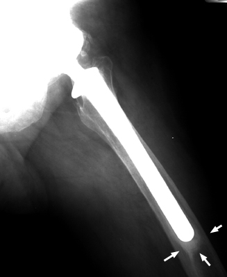
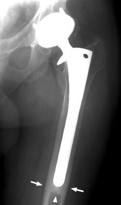
- Morphological Hallmark: The pedestal sign is characterized by a dense, transverse condensation of endosteal new bone (a neo-cortical bone bridge or shelf) spanning the femoral medullary canal immediately below the distal tip of the femoral component (Gruen Zone 4), separated from the prosthetic tip by a distinct radiolucent zone (equal to or greater than 2 mm).
- Pathognomonic Significance: In cementless total hip arthroplasty (THA), the presence of a pedestal separated by a radiolucent halo indicates failure of proximal metaphyseal osseointegration, distal load transfer, and persistent axial or angular micromotion (toggle), serving as a cardinal radiographic sign of mechanical stem loosening.
- Critical Diagnostic & Surgical Implication: Distinguishes stable biologic fixation from fibrous encapsulation or gross mechanical instability under the Engh criteria; its identification, especially alongside progressive radiolucent lines or subsidence, prompts diagnostic workup for aseptic loosening and guides preoperative planning for revision femoral reconstruction.
2. Diagnostic Sign Summary Matrix
| Primary Modality | Anatomic Location | Primary Pathology | Pathognomonic Morphology | Diagnostic Utility & Sensitivity |
|---|---|---|---|---|
| Plain Radiography (AP & Lateral Femur) | Distal Femoral Diaphysis / Gruen Zone 4 & Zone 11 | Femoral Stem Aseptic Loosening / Fibrous Encapsulation | Transverse sclerotic bone shelf across medullary canal separated from stem tip by ≥2 mm radiolucent halo | High specificity (88% to 94%) for stem instability when accompanied by subsidence or divergent radiolucencies |
| Computed Tomography (MARS-CT) | Periprosthetic Medullary Canal at Stem Tip | Distal Toggle & Fibrous Membrane Interposition | Complete transverse neocortical plate with circumferential soft-tissue attenuation (<40 HU) gap around metal tip | Superior sensitivity (96%) for circumferential lucencies, endosteal osteolysis, and neocortical bridging |
| Nuclear Medicine (SPECT-CT / Bone Scan) | Gruen Zone 4 (Femoral Tip Fulcrum) | Mechanical Interface Micromotion & Bone Remodeling | Focal, high-intensity tracer uptake concentrated exclusively at the distal tip of the femoral component | High negative predictive value (95%) for ruling out active mechanical loosening when distal uptake is absent |
3. Clinical Case Imaging & Standardized Figures
4. Pathophysiology & Biomechanical Mechanism
The development of the pedestal sign reflects fundamental biomechanical principles of load transmission, implant stability, and Wolff’s law within the reconstructed proximal femur. In an ideal cementless total hip arthroplasty, rigid initial press-fit stability allows microscopic bone ingrowth into the proximal porous coating (Gruen zones 1, 2, 6, and 7). Under stable proximal osseointegration, physiological joint reaction forces are transferred directly from the femoral head to the proximal femoral metaphysis. This physiological loading produces proximal endosteal bone remodeling—manifested as spot welds—while sparing the distal diaphysis from excessive load, leaving the medullary canal open beneath the distal tip.
Conversely, if an implant fails to achieve rigid initial press-fit or microscopic bone ingrowth—due to stem undersizing, poor host bone quality, osteopenia, or excessive early interface motion exceeding 150 micrometers—the proximal metaphyseal envelope cannot support physiological shear and compressive loads. Consequently, the stem migrates or pistons distally, transferring axial compressive loads down the length of the implant to the distal diaphyseal stem tip in Gruen Zone 4.
As the distal stem tip impacts the endosteal medullary bone during repetitive gait cycles, the localized cyclic stress concentration stimulates endosteal osteoblasts to synthesize reactive woven and lamellar bone. This endosteal osteogenesis progressively condenses into a horizontal neocortical plate across the canal, termed a pedestal, which attempts to arrest further stem subsidence. When the stem remains mechanically unstable, dynamic angular toggling and axial pistoning continually shear the interface, interposing a fibrocartilaginous or fibrous membrane between the metallic tip and the newly formed bone shelf. On radiographs, this interposed fibrous tissue manifests as a distinct radiolucent halo (equal to or greater than 2 mm) separating the prosthesis from the underlying pedestal, completing the classic pedestal sign originally characterized by Jakim and colleagues.
5. Modality-Specific Imaging Findings
Plain Radiography
High-quality, standardized anteroposterior (AP) pelvis, AP hip, and cross-table lateral or frog-leg lateral radiographs serve as the primary diagnostic foundation. Key radiographic features must be systematically analyzed according to Gruen zones (Zones 1-7 on AP, Zones 8-14 on lateral):
- Pedestal Morphology: A transverse or dome-shaped endosteal bone bridge spanning greater than 50% of the medullary canal width in Gruen Zone 4. The bone shelf displays corticated, radio-opaque margins matching cortical density.
- Radiolucent Halo: A well-defined, continuous radiolucent zone (2 mm or wider) between the metallic stem tip and the superior border of the pedestal. Parallel radiolucencies extending proximally along the stem shaft indicate progressive fibrous encapsulation.
- Cortical Hypertrophy: Thickening of the femoral cortices in Gruen zones 3, 4, and 5 reflects chronic distal load transfer and stem modulus mismatch.
- Absence of Proximal Spot Welds: Lack of endosteal new bone bridging connecting host trabecular bone to the porous-coated surface in Zones 1, 2, 6, or 7 confirms absent proximal ingrowth.
- Component Migration: Serial comparison demonstrates vertical subsidence (axial stem migration >2 mm relative to the greater trochanter or teardrop) or angular tilt into varus orientation.
Computed Tomography (MARS-CT)
Multi-detector CT with metal artifact reduction software (MARS) provides precise cross-sectional anatomic delineation of the bone-implant interface. Axial, coronal, and sagittal reconstructions confirm the complete 360-degree extent of the neocortical transverse plug. Attenuation values of the interpositional gap between the stem tip and pedestal measure soft-tissue density (20 to 40 Hounsfield Units), distinguishing fibrous tissue from fluid-filled osteolytic lesions or gas voids. Multiplanar CT also readily assesses true volumetric endosteal bone loss and detects occult periprosthetic fractures.
Magnetic Resonance Imaging & Nuclear Medicine
Optimized MARS-MRI utilizing multi-acquisition variable-resonance image combination (MAVRIC) or slice encoding for metal artifact correction (SEMAC) sequences permits detailed visualization of the periprosthetic soft-tissue envelope. MRI evaluates synovial thickness, capsular distension, and adverse local tissue reactions (ALTR) resulting from wear debris or trunnionosis. On triple-phase technetium-99m (99mTc) bone scintigraphy and single-photon emission computed tomography (SPECT-CT), mechanical stem toggle and active pedestal remodeling manifest as intense focal radiotracer uptake localized specifically to Gruen Zone 4 during delayed static phases.
6. Diagnostic Utility, Sensitivity & Specificity
In the seminal evaluation of biologic fixation by Engh, Massin, and Suthers (1990), radiographic findings were classified into three definitive categories: bone ingrowth (biologic fixation), stable fibrous fixation, and unstable (loose) fixation. The diagnostic interpretation of distal endosteal bone depends heavily on whether the bone directly contacts the stem or is separated by a lucent boundary:
- Distal Pedestal in Biologic Fixation: A true bone pedestal is absent in fully ingrown, proximally fixed stems, as proximal spot welds shield the distal diaphysis from compressive loading.
- Distal Pedestal in Stable Fibrous Fixation: A dense pedestal contacting or closely approximating the stem tip in an implant that has ceased migrating reflects stable fibrous encapsulation. The implant achieves functional load support without progressive subsidence.
- True Pedestal Sign in Unstable Stems: When a pedestal is separated from the stem tip by a radiolucent gap equal to or greater than 2 mm, or when the pedestal fails to bridge the entire canal while the stem exhibits ongoing subsidence or varus tilt, the specificity for mechanical loosening ranges from 88% to 94%.
Modern cohort investigations demonstrate that while an isolated distal pedestal on a single postoperative radiograph has moderate sensitivity (approximately 50% to 65%) for loosening, serial radiograph analysis demonstrating a newly appearing or enlarging pedestal alongside progressive radiolucent lines yields a positive predictive value exceeding 90% for clinical implant failure.
7. Differential Diagnosis & Radiographic Mimics
| Sign / Condition | Distinguishing Radiographic Features | Clinical Presentation | Key Differentiating Test |
|---|---|---|---|
| Pedestal Sign (Aseptic Loosening) | Transverse neocortex across canal separated from stem tip by ≥2 mm radiolucent halo; absent proximal spot welds; possible subsidence | Mechanical start-up thigh or groin pain on weight-bearing; progressive limp; relief with rest | Serial comparative radiographs demonstrating progressive subsidence; focal Zone 4 uptake on SPECT-CT |
| Stable Fibrous Fixation | Transverse bone pedestal closely abutting stem tip; continuous parallel radiolucent lines (<1 mm) with surrounding sclerotic halo; zero subsidence over 2 years | Mild, stable, or absent thigh pain; satisfactory functional mobility without deterioration | Serial plain radiographs showing absolute positional stability over ≥24 months; stable Engh fixation score |
| Distal Cortical Hypertrophy | Symmetric endosteal and periosteal cortical thickening in Gruen zones 3 and 5; medullary canal remains patent; proximal spot welds intact | Asymptomatic or transient stiffness; benign response to stem stiffness and modulus mismatch | Presence of robust proximal spot welds in Zones 1 and 7; absence of radiolucent lines or subsidence |
| Periprosthetic Joint Infection (PJI) | Rapidly progressive, irregular, scalloped endosteal osteolysis; patchy cortical destruction; periosteal laminar reaction; ragged radiolucencies | Constant, non-mechanical rest pain, night pain, surgical site erythema, warmth, fever, wound drainage | Serum ESR, CRP, and D-dimer; diagnostic hip aspiration with synovial fluid leukocyte count, differential, and culture |
| Retained Distal Cement Restrictor | Radio-opaque or radiolucent plastic/bone plug visible well below femoral tip; well-demarcated margins; static across all postoperative films | Asymptomatic; surgical history of prior cemented stem revision or plug placement | Operative note review confirming historical insertion of acrylic, polyethylene, or resorbable cement stop |
8. Clinical Significance & Management Implications
The clinical identification of a pedestal sign demands a comprehensive clinical and diagnostic assessment to establish whether the femoral component is clinically loose and symptomatic, or represents stable fibrous encapsulation:
- Symptomatic Mechanical Loosening: Patients with an unstable stem and a classic pedestal sign typically present with activity-related, start-up thigh pain located along the mid-to-distal anterior femur, corresponding to distal tip toggle against the endosteum. When progressive subsidence (greater than 2 to 3 mm), stem tilt into varus, or continuous divergent radiolucent lines accompany the pedestal sign, aseptic loosening is confirmed. In these symptomatic patients, conservative management is ineffective, and revision total hip arthroplasty is indicated.
- Asymptomatic Stable Fibrous Fixation: In patients who are completely asymptomatic and demonstrate no component migration across serial 12-month radiographs, conservative surveillance is indicated. These patients should be monitored with clinical examinations and standing radiographs at annual intervals to ensure osteolysis or late mechanical failure does not develop.
- Preoperative Revision Planning: The presence of a dense, sclerotic pedestal significantly impacts revision surgical technique. During stem extraction, the robust bone pedestal frequently obstructs the distal medullary canal, impeding the insertion of standard revision reamers and long-stem revision implants. The operating surgeon must plan for controlled trephination or high-speed burr removal of the pedestal, often utilizing fluoroscopic guidance or an extended trochanteric osteotomy (ETO) to prevent distal femoral cortical perforation.
- Revision Implant Selection: Because the proximal metaphyseal bone is often compromised and deficient in cases of loose cementless stems, revision implants that bypass the proximal deficiency and achieve diaphyseal scratch-fit stability—such as modular tapered fluted titanium stems or extensively porous-coated cylindrical stems—are routinely required.
9. Master Class Pro-Tip
Master Class Pro-Tip: When planning revision femoral arthroplasty in the setting of a prominent pedestal sign, never attempt to drill or ream blindly through the sclerotic bone shelf with standard rigid reamers. The dense neocortical pedestal behaves like cortical bone, readily deflecting flexible reamers or straight reamers toward the thinner anterior or lateral femoral cortex, precipitating catastrophic diaphyseal perforation. Always obtain biplanar intraoperative fluoroscopy or perform an extended trochanteric osteotomy (ETO). Direct visualization with high-speed pencil burrs allows centralized recanalization of the diaphysis, ensuring adequate reaming depth and concentric bypass for the revision diaphyseal-engaging stem.
Master Class Radiological Pearl: Femoral Canal Recanalization in Revision THA
10. Evidence-Based Synthesis
| Author & Year | Study Design | Sample Size | Key Findings & Diagnostic Accuracy | Clinical Implication | Level of Evidence | DOI |
|---|---|---|---|---|---|---|
| Jakim et al. (1989) | Case Series | N = 54 hips | Originally defined the pedestal sign as a transverse condensation of bone separated from the stem tip by ≥2 mm within a radiolucent area; established strong correlation with stem toggle and loosening. | Identifies the distal radiolucent halo and transverse bone bridge as a primary warning sign of unstable fibrous encapsulation. | Level IV | PubMed |
| Engh et al. (1990) | Comparative Cohort Study | N = 1010 hips | Established definitive criteria for biologic fixation; demonstrated that an isolated pedestal in a stable stem reflects fibrous encapsulation, whereas an incomplete pedestal or one separated by >2 mm lucency denotes instability. | Distinguishes stable fibrous osseointegration from unstable loosening based on progressive radiolucent lines and subsidence. | Level III | DOI |
| Engh et al. (1987) | Histologic & Radiographic Correlation | N = 89 specimens | Histologically demonstrated that stems with robust proximal bone ingrowth (spot welds) lack distal pedestal formation, whereas stems transferring load distally develop neocortical bone bridges. | Confirmed that distal pedestal formation is an adaptive bone response to axial micro-motion and distal load transfer away from the metaphysis. | Level IV | DOI |
| Gruen et al. (1979) | Radiographic Analysis | N = 389 hips | Defined the 7 anatomic femoral zones; identified Zone 4 (distal stem tip) as the critical biomechanical fulcrum for distal toggle, cantilever bending, and endosteal bone reaction. | Standardized the anatomical localization of distal tip cortical hypertrophy, radiolucencies, and pedestal bone bridges in Zone 4. | Level IV | DOI |
| Brown et al. (2002) | Instructional Review | Narrative Synthesis | Reviewed the pathophysiology of mid-thigh pain following cementless THA; showed that distal stem micromotion, modulus mismatch, and distal pedestal formation strongly correlate with mechanical stem toggle and pain. | Guides differential diagnosis of mid-thigh pain; presence of pedestal sign with clear radiolucent demarcation indicates mechanical instability requiring revision. | Level V | DOI |
| Bourget-Murray et al. (2024) | Prospective Cohort Study | N = 235 hips | Investigated distal bone remodeling in modern flat tapered stems; observed non-loosening pedestal formation in <2% of cases, demonstrating that stable proximal fit prevents distal load transfer and pedestal formation. | Highlights that modern short and tapered wedge stems achieve stable proximal osseointegration, making the appearance of a distal pedestal an atypical finding warranting serial follow-up. | Level IV | DOI |
| Evola et al. (2025) | Comparative Cohort Study | N = 140 hips | Compared curved short stems with straight tapered stems; confirmed that distal cortical hypertrophy and distal pedestal formation were significantly less frequent with proximal curved metaphyseal engagement. | Provides comparative evidence that proximal stem geometry governs distal bone remodeling patterns and prevents distal pedestal condensation. | Level III | DOI |
11. References
- Jakim I, Barlin C, Sweet MB. Radiological signs of loosening of the femoral stem in cementless total hip arthroplasty. S Afr J Surg. 1989;27(3):78-83. PubMed [Level IV: Case Series]
- Engh CA, Massin P, Suthers KE. Roentgenographic assessment of the biologic fixation of porous-surfaced femoral components. Clin Orthop Relat Res. 1990;(257):107-128. DOI [Level III: Comparative Cohort Study]
- Engh CA, Bobyn JD, Glassman AH. Porous-coated hip replacement. The factors governing bone ingrowth, stress shielding, and clinical results. J Bone Joint Surg Br. 1987;69(1):45-55. DOI [Level IV: Histologic & Radiographic Correlation]
- Gruen TA, McNeice GM, Amstutz HC. “Modes of failure” of cemented stem-type femoral components: a radiographic analysis of loosening. Clin Orthop Relat Res. 1979;(141):17-27. DOI [Level IV: Radiographic Analysis]
- Brown TE, Larson B, Shen F, Moskal JT. Thigh pain after cementless total hip arthroplasty: evaluation and management. J Am Acad Orthop Surg. 2002;10(6):385-392. DOI [Level V: Instructional Review]
- Bourget-Murray J, et al. Early- to mid-term outcome of a short, cementless, titanium, flat, tapered stem for primary total hip arthroplasty: an independent series. Hip Int. 2024;34(3):362-368. DOI [Level IV: Prospective Cohort Study]
- Evola FR, et al. Comparative study of fitmore and CLS stems in total hip arthroplasty: midterm clinical and radiographic outcomes. Musculoskelet Surg. 2025;109(1):45-52. DOI [Level III: Comparative Cohort Study]
Last Updated on September 28, 2026
🔗 Knowledge Graph & Clinical Correlates
OrthopaedicsOne Living Semantic Network🛠️ Operative Techniques
- Valgus osteotomy for Perthes disease Pediatrics
- Valgus osteotomy for development coxa vara Hip and Knee
🩺 Clinical Conditions & Indications
- Which EMR should I choose Original Clinical Contribution: Authored for the OrthopaedicsOne General Orthopaedics Specialty Section. Preserved, digitized, and updated to modern evidence-based...
🔬 Anatomy, Pathology & Diagnostics
- Why I Would Not Consider Navigating My THA Hip Joint & Proximal Femur
- Why I Would Consider Navigating My THA Hip Joint & Proximal Femur
- Why I Use Metal on Metal Hip Joint & Proximal Femur
- Why I Use Cross-Linked Polyethylene Hip Joint & Proximal Femur