
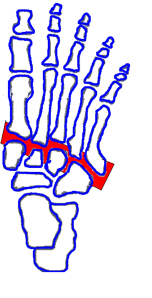
The Lisfranc joint (or complex) joins the midfoot to the forefoot. This complex comprises the articulation of the five metatarsals and the three cuneiforms medially and the cuboid laterally.

Figure 1. The red zone represents the entire articulation of the five metatarsals with the three cuneiforms and the cuboid
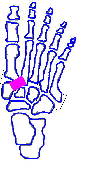
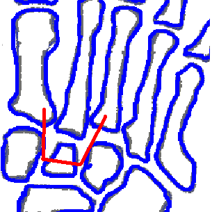
The Lisfrac joint has some inherent (bony) stability, conferred primarily by the relative posterior position of the middle cuneiform (relative to the medial and lateral cuneiforms), as shown in the figure. This provides a mortise into which the second metatarsal is recessed.
Figure 2. The intermediate cuneiform is slighted recessed relative to its neighbors, creating a mortise into which the 2nd MT can sit; this provides static stability to the medfoot-forefoot articulation
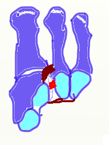
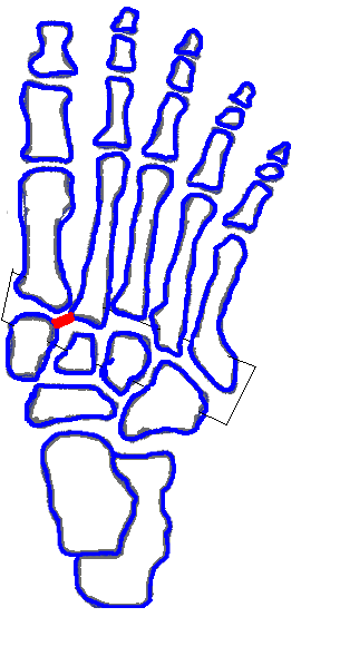
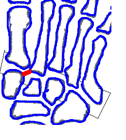
Most of the stability of the joint comes from the ligamentous attachments. These are located on both dorsal and plantar surfaces, as well as between the bones (interosseous) and course in longitudinal, oblique, and transverse directions. The so-called “Lisfrac ligament” is an oblique interosseous ligament between the medial cuneiform and second metatarsal; it is supplemented by dorsal and plantar oblique bands.
Figure 3a. The oblique interosseous ligament (red) between the medial cuneiform and the 2MT is, strictly speaking, the “Lisfranc ligament”; however, there are also dorsal and plantar bands, as shown in Figure 4c
Figure 3b. Close up view of the Lisfranc ligament
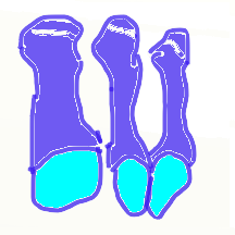
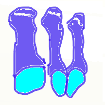
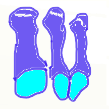
Figure 4a. This drawing shows the first, second and third metatarsals from a superior posterior position, with the midfoot removed
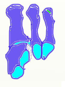
Figure 4b. In this drawing, the medial cuneiform is added; it articulates, of course, with the first metatarsal distally. The surfaces for its articulation with the navicular and intermediate cuneiform are shown.
Figure 4c. The Lisfranc ligament is shown in red; the dorsal and plantar bands are shown in rust. As seen, the dorsal band is exclusively to the 2nd MT; the plantar band attaches to the 3rd as well.
Attachments:

3mt en face.PNG (image/png)
3mt MC en face.PNG (image/png)
LL 3mt MC en face.PNG (image/png)
3mt en face.PNG (image/png)
3mt en face.PNG (image/png)
ap foot lisfranc lig dorsal.PNG (image/png)
ap foot lisfranc lig.PNG (image/png)
ap foot lisfranc.PNG (image/png)
ap foot recess for 2nd mt.PNG (image/png)
ap foot lisfranc lig close.PNG (image/png)
Last Updated on June 27, 2024
🔗 Knowledge Graph & Clinical Correlates
OrthopaedicsOne Living Semantic Network🛠️ Operative Techniques
- Weil Lesser Metatarsal Shortening Osteotomy Foot and Ankle
- Varus and Valgus Lesser Toe Deformity Correction Foot and Ankle
- Turf Toe Injuries Sports Medicine
🩺 Clinical Conditions & Indications
- Turf toe Introduction Despite its rather benign namesake, turf toe is a incapacitating injury that plagues many athletes who participate...
🔬 Anatomy, Pathology & Diagnostics
- Wrist joint Wrist and Carpus
- Vertebra prominens Surface Anatomy Lumbar Spine
- Vastus medialis Femoral Shaft
- Vastus lateralis Femoral Shaft