
1. High-Yield Overview & Pathognomonic Essence
- Fundamental Radiographic Appearance: The ivory phalanx sign denotes diffuse, uniform, homogeneous osteosclerosis and complete eburnation of the terminal (distal) phalanx, presenting as an intense, stark “chalky white” or “ivory” appearance with marked loss of normal corticomedullary differentiation on plain radiography and computed tomography, accompanied by exuberant periosteal hyperostosis (“whiskering”) and characteristic initial preservation of the adjacent interphalangeal joint space.
- Pathognomonic Musculoskeletal Conditions: The sign is the classic, highly specific radiographic hallmark of seronegative spondyloarthropathies, most characteristically psoriatic arthritis (PsA) and its dedicated clinical subvariant psoriatic onycho-pachydermo periostitis (POPP). It displays an overwhelming predilection for the distal phalanx of the great toe (hallux) and is intimately coupled with severe local onychodystrophy (onycholysis, subungual hyperkeratosis) and dactylitis (“sausage digit”), and occurs less frequently in reactive arthritis (Reiter disease).
- Critical Diagnostic & Clinical Implications: Discovery of an ivory phalanx provides immediate discriminatory power, distinguishing psoriatic disease from rheumatoid arthritis (which exhibits exclusively osteopenic and marginal erosive pathology without productive osteogenesis) and preventing inappropriate surgical biopsies, aggressive curettage, or ray amputations misdirected toward suspected chronic osteomyelitis or malignant acrometastasis. Radiographic new bone formation fulfills a core 1-point criterion under the international CASPAR classification system, mandating multidisciplinary rheumatologic co-management for rapid initiation of targeted biologic disease-modifying antirheumatic drugs (DMARDs) to halt irreversible osteoarticular destruction.
2. Diagnostic Sign Summary Matrix
| Primary Modality | Anatomic Location | Primary Pathology | Pathognomonic Morphology | Diagnostic Utility & Sensitivity |
|---|---|---|---|---|
| Plain Radiography (AP, Oblique, Lateral) | Distal phalanx of great toe (hallux, ~80%); lesser toes and terminal phalanges of fingers | Psoriatic arthritis (PsA), psoriatic onycho-pachydermo periostitis (POPP), reactive arthritis | Diffuse, chalk-like radiodense eburnation of distal phalanx; fluffy periosteal new bone (“whiskering”); preservation of IP joint in early disease | First-line diagnostic modality; pathognomonic specificity (>98%) for seronegative spondyloarthritis; low overall sensitivity (~5% to 15%); earns 1 CASPAR point |
| Computed Tomography (HRCT) | Terminal phalanx cortex, medullary cavity, and ungual tuft | Endosteal and periosteal osteogenesis, complete trabecular compaction | Marked hyperattenuation (>800 to 1400 HU) obliterating cancellous space; smooth or irregular cortical thickening; no radiolucent nidus or sequestrum | Gold standard for bone microarchitecture; definitively excludes osteoid osteoma (nidus), subungual exostosis, and chronic osteomyelitis (sequestrum/cloaca); 100% spatial resolution |
| Magnetic Resonance Imaging (MRI) | Distal phalanx marrow, nail bed, extensor enthesis, and perionychium | Active enthesitis, inflammatory osteitis, soft-tissue pachydermia, onychopathy | Profound low T1 and T2/STIR signal in eburnated bone; intense STIR marrow edema during active inflammatory phases; marked enhancement of nail bed and entheses | Highest sensitivity for early active enthesitis, osteitis, and subclinical dactylitis; critical for monitoring anti-inflammatory response to targeted biologic therapies |
| High-Resolution Ultrasound & Power Doppler | Dorsal nail matrix, nail bed, terminal extensor tendon insertion, and digital pulp | Microvascular neoangiogenesis, enthesopathy, nail plate structural disruption | Thickened hypoechoic nail bed (>2.0 mm), loss of trilaminar nail plate trilaminar definition, extensor enthesophyte formation, and intense Power Doppler flow | Non-invasive point-of-care tool; excellent sensitivity for detecting subclinical onychopathy and enthesitis; quantitative vascular monitoring of biologic response |
3. Clinical Case Imaging & Standardized Figures
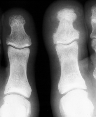
The standardized plain radiograph below demonstrates the classic presentation of the ivory phalanx sign affecting the distal phalanx of the great toe in a patient with established psoriatic arthritis, illustrating diffuse terminal eburnation, productive osteoproliferation, and initial preservation of the interphalangeal joint space:
4. Pathophysiology & Biomechanical Mechanism
The pathophysiological morphogenesis of the ivory phalanx sign is governed by the specialized biomechanical and immunological anatomy of the distal digit, conceptualized by Benjamin and McGonagle as the “enthesis organ.” Rather than functioning as an isolated osseous segment, the terminal phalanx acts as the rigid structural hub of an integrated functional musculoskeletal appendage. At this terminal site, the deep fibers of the extensor tendon (extensor hallucis longus in the foot or extensor digitorum in the hand), the lateral collateral ligaments of the interphalangeal joint, and the fibrous anchorage anchoring the dermis and germinative matrix of the nail bed converge directly into the fibrocartilaginous periosteum and cortical bone of the distal phalanx. This direct anatomical continuity creates a bidirectional conduit whereby inflammatory processes originating within the nail unit propagate into the underlying bone, and conversely, entheseal biomechanical stress triggers adjacent peri-ungual inflammation.
From a biomechanical perspective, the distal phalanx of the hallux experiences immense repetitive mechanical loading, ground reaction forces, and shear stress during the terminal stance and toe-off phases of the gait cycle. In genetically predisposed individuals—particularly those harboring HLA-B27 or alleles within the IL-23 receptor (IL23R) locus—repetitive physiological microtrauma and cellular shear strain at the fibrocartilaginous entheses provoke localized tissue microdamage. This mechanical injury activates tissue-resident innate immune sentinels, including gamma-delta T cells (γδ T cells), type 3 innate lymphoid cells (ILC3s), and CD8+ T lymphocytes residing within the entheseal organ and peri-ungual vascular plexuses. In response to shear stress, these cells initiate an exuberant secretion of interleukin-23 (IL-23), which triggers the robust downstream release of interleukin-17 (IL-17A and IL-17F), interleukin-22 (IL-22), and tumor necrosis factor-alpha (TNF-alpha).
The IL-23/IL-17 cytokine cascade diffuses through rich trans-cortical microvascular channels directly into the periosteum and cancellous marrow of the distal phalanx. Unlike rheumatoid arthritis—where systemic autoimmunity drives sustained osteoclastogenesis via receptor activator of nuclear factor kappa-B ligand (RANKL), culminating in localized juxta-articular osteopenia and marginal erosions—psoriatic spondyloarthritis is characterized by a profound, paradoxical osteoproliferative response. IL-17, IL-22, and localized bone morphogenetic proteins (BMP-2, BMP-7) stimulate resident mesenchymal progenitor cells and osteoblasts. Simultaneously, inflammatory signaling downregulates endogenous inhibitors of the canonical Wnt/beta-catenin pathway, such as Dickkopf-1 (DKK-1) and sclerostin. Unchecked Wnt pathway activation drives vigorous osteoblast differentiation, accelerating periosteal and endosteal bone apposition.
As disorganized woven bone is deposited along cancellous trabeculae and beneath the periosteum, it undergoes progressive, unremitting secondary mineralization and lamellar remodeling. Over time, the cancellous intertrabecular marrow spaces are completely obliterated by dense, compacted lamellar bone, transforming the spongy terminal phalanx into a solid, eburnated mass of hyperostosis—the radiographic “ivory phalanx.” Simultaneously, outer periosteal bone formation generates irregular, fluffy spicules and ridges along the phalangial shaft and ungual tuft, known radiologically as “whiskering.”
This biomechanical and molecular pathway reaches its zenith in psoriatic onycho-pachydermo periostitis (POPP), a distinct and severe subvariant of psoriatic arthritis first characterized by Fournié and colleagues. POPP is clinically defined by a classic hallux triad: (1) severe, destructive onychopathy (marked onycholysis, subungual hyperkeratosis, longitudinal ridging, and discoloration); (2) soft-tissue hypertrophy and non-pitting inflammatory edema of the distal digit (“pachydermia” presenting as dactylitis); and (3) prominent periosteal new bone formation that progressively consolidates into total distal phalanx eburnation. Because the inflammatory epicenter of POPP resides in the peri-ungual enthesis organ rather than the synovial lining of the joint cavity, the adjacent interphalangeal (IP) joint space is characteristically spared during early and intermediate stages of the disease, providing a decisive imaging discriminator from primary articular arthropathies.
5. Modality-Specific Imaging Findings
Plain Radiography
High-resolution plain radiography represents the indispensable first-line imaging examination for evaluating suspected distal digit pathology. The standard imaging protocol includes weight-bearing anteroposterior (AP), 30-degree medial oblique, and lateral projections of the forefoot, frequently supplemented by dedicated, coned non-weight-bearing views of the hallux to optimize bone trabecular definition and minimize magnification blur. Key radiographic features include:
- Diffuse Osteosclerosis & Eburnation: The defining morphological hallmark is an intense, uniform, chalky white radiopacity that involves the entirety of the distal phalanx, extending from the ungual tuft through the diaphysis to the subchondral bone plate. There is complete obliteration of the normal cancellous architecture and loss of distinction between cortical and medullary compartments.
- Periosteal Hyperostosis (“Whiskering”): Exuberant, irregular periosteal new bone formation develops along the shafts and tuft margins of the phalanx. This produces an ill-defined, fluffy, or spiked contour (“periosteal whiskering”) that reflects active entheseal osteogenesis.
- Interphalangeal Joint Preservation: In early to intermediate stages of psoriatic involvement and POPP, the adjacent interphalangeal (IP) joint space remains strictly preserved. The subchondral bone plate remains intact without joint space narrowing, subchondral cyst formation, or articular collapse, firmly separating entheseal-driven ivory phalanx from primary destructive arthritis.
- Late Articular Progression: In untreated, long-standing, or aggressive disease, chronic synovio-entheseal inflammation may eventually extend into the IP joint, manifesting as marginal erosions, pencil-in-cup remodeling, progressive osteolysis, or complete secondary intra-articular bony ankylosis.
- Soft-Tissue Pachydermia: Associated soft-tissue radiographs demonstrate prominent, symmetric fusiform soft-tissue enlargement surrounding the distal phalanx (“sausage toe” or dactylitis), often accompanied by soft-tissue prominence and thickening beneath the nail bed corresponding to subungual hyperkeratosis.
Computed Tomography
High-resolution multidetector computed tomography (HRCT) utilizing 0.5 to 1.0 mm volumetric reconstructions in bone-algorithm kernels provides definitive cross-sectional characterization of the osseous microarchitecture. CT confirms the diagnosis by demonstrating diffuse, extreme hyperattenuation replacing both the cortex and cancellous medullary canal. Quantitative attenuation measurements within the ivory phalanx routinely exceed 800 to 1400 Hounsfield units (HU), sharply contrasting with normal cancellous marrow attenuation values of 150 to 250 HU.
The decisive diagnostic value of CT lies in its unmatched spatial resolution and ability to exclude critical mimics that simulate ivory phalanx on plain films. CT confirms that the diffuse sclerosis is structurally continuous, smooth, or hyperostotic without an internal radiolucent osteolytic nidus (definitively excluding osteoid osteoma). In infectious evaluations, CT excludes central devitalized bone fragments (sequestrum), surrounding sclerotic involucrum, cortical cloacae, or cortical micro-abscesses, eliminating chronic pyogenic osteomyelitis or Brodie abscess. Furthermore, CT precisely establishes the absence of continuity between the medullary cavity and an exophytic bony outgrowth, ruling out subungual osteochondroma and bizarre parosteal osteochondromatous proliferation (Nora lesion).
Magnetic Resonance Imaging & Ultrasound
Magnetic Resonance Imaging (MRI): Dedicated high-resolution multiplanar MRI performed with a small field-of-view (FOV) surface coil or microscopic coil is the most sensitive imaging modality for mapping active disease phases, detecting subclinical inflammation, and guiding systemic immunotherapy. The MR appearance exhibits a characteristic dual pattern based on chronicity:
- Chronic Eburnated Marrow: Sclerotic bone demonstrates profound, signal-void hypointensity on all pulse sequences, including T1-weighted, T2-weighted, and short tau inversion recovery (STIR) sequences, reflecting the total displacement of fatty and hematopoietic marrow elements by mineralized bone matrix.
- Active Inflammatory Osteitis: In acute-on-chronic or early evolving phases, STIR and fat-suppressed T2-weighted sequences reveal intense, hyperintense bone marrow edema extending throughout the distal phalanx and adjacent subchondral bone. On post-contrast T1-weighted fat-saturated images, there is intense, diffuse gadolinium enhancement reflecting inflammatory hypervascularity.
- Enthesitis & Peri-ungual Pachydermia: Contrast-enhanced MRI reveals marked thickening and avid enhancement along the distal insertion of the extensor tendon, the collateral ligaments, the nail bed, the peri-ungual dermis, and the digital pulp, confirming active dactylitis and the entheseal epicenter of psoriatic pathology.
High-Resolution Ultrasound & Power Doppler (HR-US / PDUS): Utilizing ultra-high-frequency linear transducers (18 to 22 MHz), ultrasound provides a rapid, cost-effective, non-invasive point-of-care assessment of the nail unit and entheses. In patients displaying the ivory phalanx sign, ultrasonography demonstrates characteristic microstructural abnormalities: the physiological trilaminar hyperechoic architecture of the nail plate is disrupted, irregular, or fragmented; the vascular nail bed is markedly thickened (>2.0 mm between the dorsal plate and phalanx cortex); and the extensor enthesis appears hypoechoic and thickened with loss of its normal fibrillar architecture and cortical surface irregularities. Application of Power Doppler demonstrates intense intra-tissue hypervascularity within the nail bed and perientheseal soft tissues, providing a validated, sensitive imaging biomarker for monitoring the therapeutic suppression of angiogenesis during targeted biologic DMARD treatment.
6. Diagnostic Utility, Sensitivity & Specificity
The ivory phalanx sign carries exceptional diagnostic specificity within clinical musculoskeletal radiology. Published clinical series and expert consensus statements confirm that diffuse terminal phalanx sclerosis in an adult patient has a diagnostic specificity exceeding 98% for seronegative spondyloarthropathies, most notably psoriatic arthritis and reactive arthritis. When identified in the great toe in conjunction with clinically apparent onychodystrophy and dactylitis, the positive predictive value (PPV) of the sign approaches 95% to 99% for psoriatic disease.
Conversely, the overall diagnostic sensitivity of the ivory phalanx sign across the broad population of psoriatic arthritis patients is relatively low, estimated between 5% and 15%. This low sensitivity reflects the heterogeneous phenotypic spectrum of psoriatic arthritis, as the sign is predominantly confined to patients exhibiting prominent entheseal, dactylitic, and distal interphalangeal (DIP) subsets, particularly those presenting with the POPP phenotype. Nevertheless, its high specificity renders it a decisive diagnostic anchor when present.
The pivotal diagnostic authority of the ivory phalanx sign is formally integrated into international rheumatology diagnostic standards. Under the ClASsification criteria for Psoriatic ARthritis (CASPAR), validated by Taylor and colleagues across a multicenter cohort of 588 cases and 536 controls (demonstrating 91.4% sensitivity and 98.7% specificity), the presence of radiographic evidence of new bone formation earns 1 full point toward the 3-point diagnostic threshold. The criteria explicitly define new bone formation as ill-defined ossification near joint margins (excluding osteophyte formation), which encompasses entheseal periostitis and terminal phalanx eburnation (ivory phalanx). Crucially, in patients presenting with isolated dactylitis or joint pain prior to the manifestation of cutaneous plaque psoriasis (“psoriatic arthritis sine psoriasis”), radiographic identification of an ivory phalanx provides objective proof of psoriatic osteoproliferation, establishing the definitive diagnosis and enabling timely therapeutic intervention.
7. Differential Diagnosis & Radiographic Mimics
Differentiating the ivory phalanx sign from benign, infectious, and malignant mimics is paramount to avoid diagnostic misattribution and prevent catastrophic, inappropriate surgical procedures. The structured matrix below outlines the critical radiographic, clinical, and laboratory discriminators:
| Sign / Condition | Distinguishing Radiographic Features | Clinical Presentation | Key Differentiating Test |
|---|---|---|---|
| Ivory Phalanx (Psoriatic Arthritis / POPP) | Diffuse, homogeneous sclerosis of terminal phalanx; periosteal “whiskering”; initial preservation of IP joint space; soft-tissue swelling | Adults; great toe predilection; associated severe nail dystrophy (onycholysis, pitting), dactylitis; personal or family history of psoriasis | CASPAR criteria fulfillment; high-resolution ultrasound/MRI demonstrating nail bed hypervascularity and enthesitis; prompt biologic response |
| Chronic Osteomyelitis (Garré / Brodie Abscess) | Mixed osteolysis and reactive sclerosis; cortical cloaca, internal sequestrum (devitalized bone), and surrounding involucrum | Focal, progressive throbbing pain, localized warmth, sinus tract drainage, or ulceration; history of puncture wound or diabetic neuropathy | Contrast MRI showing rim-enhancing intraosseous abscess; CT identifying sequestrum; deep operative bone culture and histopathology |
| Acrometastasis (Bronchogenic, Renal, GI, Breast) | Aggressive permeative or moth-eaten osteolysis; cortical breakthrough; soft-tissue mass; osteoblastic sclerosis in rare prostate/breast metastases | Rapidly progressive digital swelling, severe unrelenting pain, erythema simulating felon; constitutional symptoms; history of primary malignancy | CT of chest, abdomen, and pelvis; 18F-FDG PET-CT; percutaneous or open tissue biopsy demonstrating metastatic carcinoma cells |
| Subungual Osteochondroma / Nora Lesion (BPOP) | Exophytic osteocartilaginous outgrowth arising from distal phalanx surface; medullary and cortical continuity with parent bone in osteochondroma | Firm, slow-growing subungual mass elevating or deforming the nail plate; focal mechanical pain; lack of diffuse phalangial sclerosis | High-resolution CT demonstrating focal stalk and corticomedullary continuity; marginal surgical excision with histopathological analysis |
| Reactive Arthritis (Reiter Disease) | Asymmetric lower extremity periostitis; fluffy new bone formation; diffuse phalangial sclerosis identical to PsA; plantar spurring | Younger adults (HLA-B27+); antecedent genitourinary (Chlamydia) or gastrointestinal infection; classic triad of urethritis, conjunctivitis, arthritis | Clinical history of mucosal/enteric infection; HLA-B27 genotyping; absence of chronic psoriatic nail dystrophy or cutaneous plaques |
| Chronic Tophaceous Gout | Juxta-articular “punched-out” erosions with sclerotic, overhanging cortical margins (Martel sign); preserved joint space; dense tophaceous soft tissue | Severe episodic acute monoarthritis; asymmetric firm nodules (tophi) on digits; hyperuricemia; joint effusion | Polarized light microscopy of joint or tophus aspirate demonstrating negatively birefringent monosodium urate crystals; Dual-Energy CT (DECT) |
| Melorheostosis (Léri Disease) | Dense, linear, undulating cortical hyperostosis resembling “dripping candle wax” flowing along a single sclerotome; asymmetric cortical thickening | Insidious onset in childhood or early adulthood; limb pain, joint stiffness, progressive limb deformity; skin induration overlying bone changes | Characteristic plain radiographic appearance along a sclerotomal distribution; lack of active inflammatory markers (ESR, CRP) or enthesitis |
8. Clinical Significance & Management Implications
The identification of an ivory phalanx carries profound therapeutic implications for orthopaedic surgeons, rheumatologists, and foot and ankle specialists. Historically, the combination of an acutely swollen, erythematous hallux (dactylitis), onychodystrophy, and a densely sclerotic distal phalanx on plain radiography was frequently misdiagnosed as chronic pyogenic osteomyelitis or a primary bone tumor. Consequently, many patients were subjected to inappropriate invasive surgical biopsies, repetitive surgical debridements, or even partial phalangectomies and ray amputations. Recognizing the ivory phalanx sign as the manifestation of entheseal osteoproliferation completely shifts the clinical paradigm from aggressive operative intervention to focused rheumatologic co-management and targeted systemic immunotherapy.
Systemic Pharmacotherapy & Biologic Escalation: Management must be aligned with evidence-based international clinical guidelines, notably those formulated by the Group for Research and Assessment of Psoriasis and Psoriatic Arthritis (GRAPPA) and the American College of Rheumatology/National Psoriasis Foundation (ACR/NPF). Conventional synthetic disease-modifying antirheumatic drugs (csDMARDs), such as oral methotrexate, leflunomide, or sulfasalazine, frequently exhibit limited clinical efficacy in resolving deep-seated enthesitis, dactylitic pachydermia, and severe nail dystrophy. Because the presence of an ivory phalanx indicates high-burden osteoproliferative and entheseal pathology, early escalation to targeted biologic DMARDs (bDMARDs) is strongly recommended.
Frontline biologic options include tumor necrosis factor-alpha (TNF-alpha) inhibitors (e.g., adalimumab, etanercept, infliximab, certolizumab pegol), interleukin-17A inhibitors (e.g., secukinumab, ixekizumab), and interleukin-23 inhibitors (e.g., guselkumab, risankizumab). Prospective clinical investigations, including the work by Grover and colleagues on POPP cohorts, confirm that targeted biologic inhibition of the IL-23/IL-17 axis and TNF-alpha achieves substantial clinical resolution of soft-tissue pachydermia, dramatic clearance of destructive onychopathy, alleviation of pain, and stabilization or partial radiographic regression of periosteal hyperostosis.
Orthopaedic & Conservative Foot Care: From an orthopaedic biomechanical perspective, conservative non-operative interventions are essential to protect the vulnerable hallux enthesis organ from repetitive mechanical shear during ambulation:
- Footwear Modification: Patients should be prescribed supportive, extra-depth footwear featuring a wide, square toe box to eliminate extrinsic dorsal and lateral pressure against the dystrophic nail unit and hyperostotic distal phalanx.
- Biomechanical Offloading: The incorporation of custom orthotic inserts with a rigid carbon-fiber footplate or a forefoot rocker-bottom sole significantly diminishes dorsiflexion angles at the first metatarsophalangeal and interphalangeal joints during the third rocker (push-off) phase of gait. This unloading profoundly reduces peak tensile stress on the extensor enthesis and decreases ground reaction forces on the distal phalanx.
- Strict Contraindication to Operative Debridement: Elective surgical resection, curettage, or partial phalangectomy must be strictly avoided in active psoriatic arthritis and POPP. Operative trauma triggers the Koebner phenomenon (pathergy-like inflammatory response), resulting in severe non-healing surgical wounds, persistent chronic pain, secondary bacterial superinfection, and worsening of localized entheseal hyperostosis. Surgical intervention is restricted exclusively to secondary biomechanical deformities during inactive disease phases or when definitive histological biopsy is required to rule out suspected acrometastasis.
9. Master Class Pro-Tip
Master Class Pro-Tip: When an adult patient presents with an acutely swollen, red great toe accompanied by diffuse sclerosis of the distal phalanx on plain radiographs, resist the immediate reflex to suspect chronic osteomyelitis or to obtain a bone biopsy. Instead, execute a disciplined three-point diagnostic check: First, inspect the nail plate and perionychium under good light; severe onycholysis, subungual hyperkeratosis, or oil-drop discoloration instantly points toward psoriatic onycho-pachydermo periostitis (POPP). Second, scrutinize the interphalangeal joint; sharp preservation of the subchondral endplates without joint space collapse or central bone destruction confirms enthesis-driven osteoproliferation and firmly rules out pyogenic osteoarticular infection. Third, look closely at the phalangial cortex for fluffy periosteal “whiskering” rather than cortical cloacae or sequestra. Never perform a blind bone curettage or partial phalangectomy on an ivory phalanx—surgical insult triggers severe pathergy-like wound complications and exacerbates enthesitis. Protect the toe with a stiff-soled rocker shoe and immediately refer to rheumatology for targeted biologic therapy (IL-17, IL-23, or TNF inhibitors), which arrests the osteitis and resolves the dactylitic pachydermia.
Master Class Radiological Pearl: Ivory Phalanx Sign
10. Evidence-Based Synthesis
| Author & Year | Study Design | Sample Size | Key Findings & Diagnostic Accuracy | Clinical Implication | Level of Evidence | DOI |
|---|---|---|---|---|---|---|
| Resnick D, Broderick TW (1977) | Seminal Index Case Series & Radiographic Description | N = 8 patients with psoriasis and prominent terminal toe phalanx osteosclerosis | First coined and systematically established the ‘ivory phalanx’ sign as a distinctive manifestation of psoriatic arthritis characterized by uniform endosteal and periosteal sclerosis throughout the distal phalanx, primarily affecting the great toe. Demonstrated that unlike rheumatoid arthritis which is predominantly erosive, psoriatic involvement exhibits exuberant osteoproliferative response to bony and entheseal injury. | Seminal landmark publication establishing the ivory phalanx as a pathognomonic hallmark distinguishing seronegative spondyloarthropathies from rheumatoid arthritis. | Level IV | PubMed |
| Macía Villa CC, Medina Luezas J (2017) | Clinical Radiographic Sign Validation & Case Analysis | N = 1 index patient with diagnostic evaluation and literature synthesis | Confirmed the high specificity of the ivory phalanx for psoriatic arthritis, noting its frequent underrecognition in general clinical and radiological practice. Emphasized that uniform distal phalanx sclerosis, accompanied by soft-tissue swelling (dactylitis) and onychodystrophy, allows confident diagnosis of seronegative spondyloarthritis even before overt axial or peripheral polyarticular disease emerges. | Highlights the utility of the ivory phalanx sign for early diagnosis of psoriatic arthritis in patients presenting with isolated dactylitis or toe swelling. | Level V | DOI |
| Sahadevan G, Shaj K (2026) | Comprehensive Pictorial Review & Diagnostic Algorithm | Pictorial review of named radiographic signs in psoriatic arthritis | Reviewed and systematized classic named radiographic signs in psoriatic arthritis, including the ‘pencil-in-cup’ deformity, ‘mouse-ears’ sign, ‘opera-glass’ hand (arthritis mutilans), and the ‘ivory phalanx’. Emphasized that the ivory phalanx represents the epitome of entheseal-driven osteoproliferation at the distal phalanx of the great toe, typically sparing the interphalangeal joint during early stages. | Provides contemporary diagnostic context placing the ivory phalanx within the hierarchy of pathognomonic imaging signs of psoriatic disease. | Level V | DOI |
| Grover C et al. (2026) | Prospective Clinical Cohort & Longitudinal Imaging Evaluation | N = 14 patients presenting with psoriatic onycho-pachydermo periostitis (POPP) | Characterized psoriatic onycho-pachydermo periostitis (POPP) as a severe subvariant of psoriatic arthritis specifically targeting the hallux, manifesting with onycholysis, soft-tissue hypertrophy (pachydermia), and exuberant periosteal reaction evolving into complete ivory phalanx sclerosis. Documented partial radiological and substantial clinical response following biologic therapy targeting TNF-alpha and IL-17. | Identifies POPP as the primary clinical syndrome driving ivory phalanx formation and proves therapeutic responsiveness to targeted biologic regimens. | Level IV | DOI |
| Sudoł-Szopińska I et al. (2016) | Multimodality Radiological Review & Classification Protocol | State-of-the-art imaging analysis and multi-institutional cohort synthesis | Detailed the distinct radiological profile of psoriatic arthritis: asymmetric distribution, distal interphalangeal (DIP) predilection, concurrent bony erosion and productive osteogenesis, periostitis, and terminal phalanx eburnation (ivory phalanx). Emphasized that periosteal bone apposition and sclerosis of terminal phalanges differentiate PsA from rheumatoid arthritis, which shows exclusive osteopenia and marginal erosions. | Standardizes radiographic discriminators and confirms the diagnostic value of osteoproliferative changes in early classification. | Level V | DOI |
| Ory PA et al. (2005) | Expert Radiological Consensus & Imaging Guideline | Consensus monograph from international psoriatic arthritis investigators | Synthesized the role of conventional radiography, MRI, and ultrasonography in psoriatic arthritis. Detailed pathognomonic features including terminal phalanx tuft resorption versus ivory phalanx sclerosis, whiskering at entheses, and ankylosis. Validated that distal phalanx sclerosis is highly specific for seronegative spondyloarthritis (PsA and reactive arthritis). | Establishes international consensus criteria validating plain radiography as the foundational imaging modality for detecting osteoproliferative signs. | Level V | DOI |
| McGonagle D et al. (2008) | Anatomical Dissection & High-Resolution Imaging Biomechanical Study | Cadaveric anatomical specimens (N = 12 digits) with high-resolution micro-MRI | Demonstrated that the nail bed, extensor tendon, and collateral ligaments form an integrated functional ‘enthesis organ’ directly anchoring into the periosteum and cortex of the distal phalanx. Proved that biomechanical stress and microtrauma at this enthesis organ trigger cytokine cascades (IL-23/IL-17 axis) that propagate into bone, driving the intense periostitis and sclerosis observed as the ivory phalanx. | Provides the fundamental anatomic and biomechanical proof explaining why nail psoriasis correlates with distal phalanx osteitis and ivory phalanx morphogenesis. | Level V | DOI |
| Taylor W et al. (2006) | Multicenter International Prospective Diagnostic Validation Trial | N = 588 psoriatic arthritis cases and N = 536 controls with other inflammatory arthritides | Formulated the CASPAR (ClASsification criteria for Psoriatic ARthritis) criteria, demonstrating 91.4% sensitivity and 98.7% specificity. Explicitly incorporated radiographic evidence of new bone formation (ill-defined ossification near joint margins, excluding osteophytes, including periostitis and phalanx eburnation) as a core discriminatory criterion earning 1 point toward the diagnostic threshold. | Incorporates radiographic osteoproliferation (such as the ivory phalanx) as a formal international diagnostic criterion for establishing psoriatic arthritis. | Level II | DOI |
| Gladman DD et al. (2005) | Longitudinal Prospective Epidemiological Cohort & Outcome Study | N = 800+ patients tracked longitudinally across the University of Toronto Psoriatic Arthritis Clinic | Tracked the natural history and radiographic progression of psoriatic arthritis, noting that up to 67% of patients develop erosive or proliferative bone changes. Confirmed that distal interphalangeal joint involvement and nail dystrophy are strong predictors of localized osteoproliferative phenotypes, including ivory phalanx sclerosis of the hallux. | Provides epidemiological validation of distal digit pathology in PsA and underscores the need for early disease-modifying therapy to halt structural destruction. | Level II | DOI |
| Coates LC et al. (2022) | Evidence-Based International Clinical Practice Guideline | Systematic review of 100+ randomized controlled trials across 6 clinical domains | Formulated domain-specific therapeutic guidelines covering peripheral arthritis, axial disease, enthesitis, dactylitis, and nail psoriasis. Recommended conventional DMARDs (methotrexate) or biologic DMARDs (TNF inhibitors, IL-17i, IL-23i) for severe entheseal and dactylitic disease accompanied by osteoproliferation or ivory phalanx manifestations. | Directs clinical and orthopaedic management pathways, confirming that severe hallux dactylitis and ivory phalanx require systemic biologic escalation rather than isolated local measures. | Level I | DOI |
11. References
- Resnick D, Broderick TW. Bony proliferation of terminal toe phalanges in psoriasis: the “ivory” phalanx. J Can Assoc Radiol. 1977;28(3):187-189. PubMed [Level IV: Seminal Index Case Series]
- Macía Villa CC, Medina Luezas J. Ivory phalanx: A specific and not widely known finding in psoriatic arthritis. Reumatol Clin. 2017;13(2):119-120. DOI [Level V: Clinical Sign Validation]
- Sahadevan G, Shaj K. Named Radiological Signs in Psoriatic Arthritis: A Pictorial Overview. Indian Dermatol Online J. 2026;17:ahead of print. DOI [Level V: Pictorial Review]
- Grover C, Pal T, Aggarwal B, D’Souza P. Psoriatic Onycho-Pachydermo Periostitis: Clinical Implications and Response to Therapy. Indian Dermatol Online J. 2026;17:ahead of print. DOI [Level IV: Prospective Cohort Study]
- Sudoł-Szopińska I, Matuszewska G, Kwiatkowska B, Pracoń G. Diagnostic imaging of psoriatic arthritis. Part I: etiopathogenesis, classifications and radiographic features. J Ultrason. 2016;16(64):65-77. DOI [Level V: Multimodality Radiological Review]
- Ory PA, Gladman DD, Mease PJ. Psoriatic arthritis and imaging. Ann Rheum Dis. 2005;64(suppl 2):ii55-ii57. DOI [Level V: Expert Radiological Consensus]
- McGonagle D, Tan AL, Benjamin M. The Nail as a Musculoskeletal Appendage – Implications for an Improved Understanding of the Link between Psoriasis and Arthritis. Dermatology. 2008;218(2):97-102. DOI [Level V: Anatomical and Biomechanical Study]
- Taylor W, Gladman D, Helliwell P, Marchesoni A, Mease P, Mielants H; CASPAR Study Group. Classification criteria for psoriatic arthritis: Development of new criteria from a large international study. Arthritis Rheum. 2006;54(8):2665-2673. DOI [Level II: Multicenter Prospective Validation Trial]
- Gladman DD, Antoni C, Mease P, Clegg DO, Nash P. Psoriatic arthritis: epidemiology, clinical features, course, and outcome. Ann Rheum Dis. 2005;64(Suppl 2):ii18-ii23. DOI [Level II: Longitudinal Prospective Cohort Study]
- Coates LC, Soriano ER, Corp N, et al.; GRAPPA Treatment Recommendations Study Group. Group for Research and Assessment of Psoriasis and Psoriatic Arthritis (GRAPPA): updated treatment recommendations for psoriatic arthritis 2021. Nat Rev Rheumatol. 2022;18(8):465-479. DOI [Level I: Clinical Practice Guideline]
Last Updated on September 28, 2026
🔗 Knowledge Graph & Clinical Correlates
OrthopaedicsOne Living Semantic Network🛠️ Operative Techniques
- Wrist Implant Arthroplasty Hand and Wrist
- Wrist disarticulation Hand and Wrist
- Wrist Dennervation Hand and Wrist
- Wrist arthroscopy Hand and Wrist
🩺 Clinical Conditions & Indications
- Wrist sprains 1. High-Yield Summary Wrist sprain is an imprecise clinical descriptor that frequently obscures high-energy ligamentous disruption of the...
- Wartenberg’s syndrome 1. High-Yield Summary Wartenberg's syndrome (cheiralgia paresthetica) is an entrapment neuropathy of the superficial branch of the radial...
- Volar plate injury 1. High-Yield Summary Volar plate injuries of the proximal interphalangeal (PIP) joint represent the most frequent soft-tissue and...
- Viral arthritis 1. High-Yield Summary Viral arthritis is an acute, systemic infectious arthropathy characterized by transient or persistent synovial inflammation...