
1. High-Yield Overview & Pathognomonic Essence
The heel pad sign represents an objective radiographic marker reflecting systemic soft tissue and fibro-adipose expansion:
- Morphologic & Radiographic Appearance: Diffuse, symmetrical thickening of the subcalcaneal soft tissue mantle on non-weightbearing lateral foot radiographs, defined by a shortest vertical distance between the inferior-most cortical margin of the calcaneal tuberosity and the plantar skin interface exceeding 23 mm (or greater than 21.5 mm in adult females and greater than 25 mm in non-obese populations).
- Pathognomonic Pathology: The classical peripheral musculoskeletal manifestation of adult growth hormone (GH) hypersecretion and elevated serum insulin-like growth factor 1 (IGF-1) in acromegaly, producing unconstrained fibroblastic proliferation, extracellular mucopolysaccharide accumulation, and diffuse fibro-adipose hypertrophy.
- Diagnostic Utility & Clinical Implication: Provides an accessible, non-invasive plain radiographic screening indicator that can unmask insidious pituitary disease years before overt systemic, cardiovascular, or craniomaxillofacial complications emerge; however, because post-treatment fibrous remodeling resolves very slowly, it remains an insensitive index of acute biochemical disease remission.
2. Diagnostic Sign Summary Matrix
| Primary Modality | Anatomic Location | Primary Pathology | Pathognomonic Morphology | Diagnostic Utility & Sensitivity |
|---|---|---|---|---|
| Plain Radiography (Non-weightbearing Lateral Hindfoot) | Plantar subcalcaneal soft tissue space (calcaneal tuberosity to plantar skin margin) | Acromegaly (growth hormone hypersecretion / IGF-1 excess) | Shortest distance between the inferior calcaneal cortex and plantar skin surface > 23 mm (weight- and sex-adjusted) | Primary initial screening modality; sensitivity 80% to 85%, specificity 92% to 96% when adjusted for body weight at standard 40-inch source-to-image distance. |
| High-Resolution Musculoskeletal Ultrasound | Plantar heel pad fibro-adipose composite and plantar aponeurosis | Generalized fibro-adipose expansion and non-inflammatory matrix hypertrophy | Uniform vertical expansion of uncompressed fat pad (> 21 to 23 mm) with preservation of organized hyperechoic septal microchambers | Rapid, radiation-free bedside diagnostic validation; strong correlation with lateral radiography (r = 0.94); clearly differentiates generalized soft tissue expansion from focal plantar fasciitis. |
| Computed Tomography (CT Hindfoot) | Calcaneal tuberosity, subcalcaneal fat mantle, and plantar entheses | Acral osseous apposition and diffuse soft tissue overgrowth | Symmetric soft tissue thickening > 23 mm maintaining normal adipose attenuation (-80 to -120 HU), accompanied by calcaneal enthesophytes and cortical hyperostosis | Definitive 3D anatomical quantification; eliminates radiographic magnification distortion; rules out occult calcaneal fractures, destructive osteomyelitis, and soft tissue neoplasia. |
| Magnetic Resonance Imaging (MRI Hindfoot) | Subcutaneous fat pad, plantar aponeurosis, and intrinsic pedal musculature | Fibro-adipose hypertrophy without acute inflammatory fluid or edema | Diffuse expansion of plantar fat pad showing homogeneous T1 hyperintensity and intermediate T2 signal without abnormal STIR hyperintensity or contrast enhancement | High specificity (> 95%); reliably excludes acute calcaneal contusion, plantar fascial rupture, infective cellulitis/abscess, and inflammatory enthesitis associated with seronegative spondyloarthropathies. |
3. Clinical Case Imaging & Standardized Figures
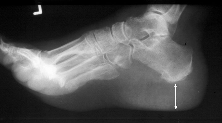
Figure 1 illustrates the classic radiographic manifestation of the heel pad sign on a standardized non-weightbearing lateral projection of the hindfoot. Measuring the shortest vertical distance between the inferior-most cortical margin of the calcaneal tuberosity and the outer plantar skin boundary reveals profound soft tissue widening measuring well in excess of the normal 21 mm threshold. The soft tissue envelope demonstrates uniform, diffuse expansion without focal lobular disruption, fluid layering, or architectural effacement. The underlying calcaneal trabecular architecture remains intact without cortical violation or fracture lines, highlighting that the expansion represents chronic endocrine-driven fibro-adipose deposition rather than acute traumatic edema or hematoma.
4. Pathophysiology & Biomechanical Mechanism
The heel pad sign is the peripheral soft tissue manifestation of prolonged, unmitigated somatotropic axis activation. In adult acromegaly, a benign somatotroph adenoma of the anterior pituitary gland autonomously hypersecretes growth hormone (GH), which stimulates hepatic and peripheral production of insulin-like growth factor 1 (IGF-1). Because physical growth plates (physes) undergo physiologic fusion at the cessation of puberty, longitudinal skeletal growth is no longer possible. Instead, persistent GH and IGF-1 elevation acts upon receptive mesenchymal cells, periosteal osteoblasts, synovial chondrocytes, and subcutaneous fibroblasts throughout acral anatomic sites.
The biochemical and cellular mechanisms driving calcaneal heel pad expansion comprise three coordinated pathophysiological processes:
- Extracellular Glycosaminoglycan Deposition & Interstitial Hydration: IGF-1 binds to transmembrane IGF-1 receptors on dermal and subcutaneous fibroblasts, activating intracellular MAPK and PI3K-Akt signaling cascades. This markedly upregulates the transcription and secretion of core extracellular matrix proteins and negatively charged glycosaminoglycans (GAGs), principally hyaluronic acid and chondroitin sulfate. Owing to their high polyanionic charge density, GAGs avidly bind mobile cations and water molecules, creating high osmotic swelling pressure and dramatic hydrophilic expansion within the interstitial matrix.
- Fibro-Adipose Hyperplasia & Architecture Maintenance: The calcaneal fat pad is a specialized mechanical organ engineered for shock attenuation, composed of enclosed hydraulic microchambers and macrochambers bounded by dense, spiraling fibro-elastic septae anchored from the calcaneal periosteum to the deep reticular dermis. Under prolonged GH stimulation, adipocytes undergo both hypertrophy and preadipocyte recruitment (hyperplasia), while the fibrous septae undergo thickened collagenous remodeling. Unlike traumatic septal rupture or degenerative fat pad atrophy, the acromegalic heel pad preserves its internal compartmentalized structural architecture while its total tissue volume expands symmetrically.
- Periosteal Appositional Hyperostosis & Enthesopathy: GH and IGF-1 directly stimulate osteoblasts within the vascular periosteum, initiating appositional new bone formation. At high-stress mechanical attachment sites, traction enthesophytes develop at the calcaneal tuberosity along the origin of the central plantar aponeurosis and the insertion of the Achilles tendon. Concurrent subchondral bone remodeling and cartilage hypertrophy across adjacent tarsal articulations alter biomechanical force transmission during gait.
From a biomechanical standpoint, the hypertrophied acromegalic heel pad exhibits altered visco-elastic damping properties. Although tissue volume is substantially expanded, collagen cross-linking and proteoglycan overloading increase internal matrix stiffness. As a result, energy dissipation during initial ground contact is impaired, transferring increased repetitive impact stresses across the subtalar joint, midtarsal joints, and metatarsophalangeal articulations, accelerating secondary degenerative osteoarthritis.
5. Modality-Specific Imaging Findings
Plain Radiography
Non-weightbearing lateral radiography of the foot remains the historic benchmark and primary screening tool for measuring heel pad thickness. Standardized acquisition mandates a 40-inch (100 cm) source-to-image distance (SID) centered over the hindfoot to avoid geometric beam divergence and false magnification. Measurement is performed by identifying the shortest vertical line connecting the inferior-most cortical contour of the calcaneus to the radiolucent skin margin on the plantar aspect of the heel.
Normal heel pad thickness averages 17.8 mm in healthy females and 19.5 mm in healthy males. Radiographic heel pad thickness exceeding 21.5 mm in adult women and 23.0 mm in adult men warrants suspicion, while values exceeding 25.0 mm in non-obese individuals are highly specific for acromegaly. Secondary osseous findings readily visible on plain radiographs include prominent plantar calcaneal spurs, hypertrophic Achilles enthesophytes, terminal tuft mushrooming of the distal phalanges (“spade-like” phalanges), widening of the joint spaces secondary to chondral hyperplasia in early stages, and prominent cortical hyperostosis across the metatarsal shafts.
Computed Tomography
Computed tomography (CT) provides multiplanar volumetric reconstructions that eliminate optical projection parallax and magnification artifacts inherent to plain films. On sagittal and coronal high-resolution CT reconstructions, heel pad thickness can be measured with sub-millimeter precision. The subcalcaneal soft tissues demonstrate homogeneous low attenuation characteristic of mature adipose tissue (-80 to -120 Hounsfield units), interspersed with delicate, regular soft tissue attenuation bands (+10 to +30 HU) representing intact fibro-elastic septae.
CT excels at evaluating the underlying osseous framework, confirming the absence of subtle calcaneal stress fractures or cortical erosions, detailing the exact morphology and size of plantar calcaneal spurs, and characterizing secondary osteoarthritic joint space narrowing, subchondral sclerosis, and osteophytosis in advanced acromegalic arthropathy.
Magnetic Resonance Imaging & Ultrasound
Magnetic resonance imaging (MRI) provides superior contrast resolution of the soft tissue compartments of the hindfoot. In patients with acromegaly, sagittal T1-weighted and T2-weighted fast spin-echo images demonstrate marked expansion of the subcutaneous fat pad with preservation of normal adipose signal intensity: high signal on non-fat-suppressed T1 sequences and intermediate signal on T2 sequences. Short tau inversion recovery (STIR) and T2-weighted fat-saturated sequences demonstrate complete absence of abnormal fluid signal, hyperintensity, or diffuse reticulation within the fat pad, definitively ruling out acute traumatic contusion, inflammatory cellulitis, or systemic interstitial edema. The proximal plantar aponeurosis typically maintains a normal thickness of less than 4.0 mm, confirming that the expansion is not driven by localized plantar fasciitis.
High-resolution musculoskeletal ultrasound (7.5 to 15 MHz linear array) represents a rapid, cost-effective, radiation-free diagnostic modality. With the patient prone and the foot hanging freely in a non-weightbearing state, the transducer is positioned longitudinally along the plantar midline of the calcaneal tuberosity without applying downward transducer pressure. Ultrasound directly measures the uncompressed vertical distance between the bright hyperechoic calcaneal bony reflection and the superficial epidermal margin. Ultrasound demonstrates intact, hyperechoic fibro-elastic septal architecture bounding hypoechoic fat lobules. Color Doppler evaluation reveals no internal hypervascularity, differentiating acromegalic expansion from active inflammatory enthesopathies or soft tissue neoplasms.
6. Diagnostic Utility, Sensitivity & Specificity
The diagnostic accuracy of heel pad thickness as a surrogate marker for acromegaly has been extensively validated in historic and contemporary cohorts:
- Sensitivity: Across landmark radiographic series (Steinbach & Russell 1964; Kho et al. 1970; MacSweeney et al. 1990), using a standard threshold of > 23 mm yields a diagnostic sensitivity ranging between 80% and 85% for acromegalic patients.
- Specificity: When adjusted for patient sex and body mass, specificity reaches 92% to 96%. Setting a conservative diagnostic cutoff of > 25 mm elevates specificity above 98% in non-obese clinical cohorts.
- Positive & Negative Predictive Values: In general outpatient populations with undifferentiated foot pain, the positive predictive value (PPV) is moderated by the prevalence of obesity and dependent edema. However, in patients presenting with concomitant acral symptoms (enlarging shoe or ring size, carpal tunnel syndrome, coarse facial skin, macroglossia), a heel pad exceeding 23 mm carries a PPV surpassing 90%, necessitating immediate endocrine referral. The negative predictive value (NPV) is approximately 88%, as a normal heel pad measurement does not completely exclude early or mild biochemical acromegaly.
Several technical and physiological confounding factors influence measurement precision and must be controlled:
- Patient Body Weight and Body Mass Index (BMI): Body weight exerts a direct physiological influence on soft tissue thickness. Kho et al. established that heel pad thickness increases by approximately 1 mm for every 10 to 15 kg increase in body weight. In morbidly obese individuals, heel pad thickness can exceed 23 mm purely secondary to adipose loading, requiring correlation with biochemical assays.
- Weightbearing vs Non-weightbearing Radiography: Upright weightbearing compresses the hydraulic chambers of the fat pad, forcing the soft tissues to spread laterally and reducing the vertical measurement by 2 to 4 mm. Diagnostic thresholds are strictly standardized on non-weightbearing lateral projections.
- Geometric Magnification & Source-to-Image Distance (SID): Standard tables are calibrated at a 40-inch (100 cm) SID. Reducing the focal distance to 36 inches or improper positioning of the hindfoot away from the detector amplifies geometric divergence, falsely increasing apparent soft tissue thickness by 1.5 to 2.5 mm.
7. Differential Diagnosis & Radiographic Mimics
| Sign / Condition | Distinguishing Radiographic Features | Clinical Presentation | Key Differentiating Test |
|---|---|---|---|
| Acromegaly (Classic Heel Pad Sign) | Diffuse, symmetrical soft tissue thickening > 23-25 mm; normal fat attenuation/signal; calcaneal spurs; phalangeal tuft mushrooming; joint widening | Insidious shoe/ring size increase, facial coarsening, macroglossia, hyperhidrosis, carpal tunnel syndrome, arthralgias | Serum IGF-1 (elevated) and Oral Glucose Tolerance Test (OGTT) with failure of GH suppression; pituitary MRI |
| Obesity / Increased Body Weight | Symmetrical heel pad thickening (typically 21-24 mm) proportional to BMI; lateral spread on weightbearing; absence of acral tufting | Elevated BMI, absence of somatic acral enlargement or facial changes, mechanical heel pain | Normal serum IGF-1; suppressed GH on testing; normal pituitary imaging |
| Plantar Fasciitis | Localized fusiform thickening of proximal plantar fascia origin (> 4.0-4.5 mm) with surrounding perivascular fat stranding; diffuse heel pad thickness normal (< 21 mm) | “First-step” morning heel pain, focal tenderness at medial calcaneal tubercle; insidious onset | High-resolution ultrasound or MRI showing proximal plantar fascia thickening, intrasubstance hyperintensity, and calcaneal bone marrow edema |
| Calcaneal Contusion / Acute Trauma | Acute focal or diffuse soft tissue swelling with loss of clear septal architecture, soft tissue hematoma, possible trabecular microfractures or cortical disruption | Acute history of high-energy impact/fall from height; severe localized pain, ecchymosis, inability to bear weight | CT / MRI showing calcaneal trabecular impaction/fracture and extensive T2/STIR fat pad edema/hemorrhage |
| Peripheral Edema (CHF, Renal Failure, Venous Stasis) | Diffuse subcutaneous thickening with reticular soft tissue stranding, fluid layering, and ill-defined skin-subcutaneous interface; bilateral | Dependent, pitting edema of ankles and feet, dyspnea, jugular venous distension, stasis dermatitis | Clinical examination (pitting), echocardiography, renal function panels, venous duplex ultrasound; resolves with limb elevation and diuresis |
| Myxedema / Severe Hypothyroidism | Moderate diffuse soft tissue thickening secondary to dermal and interstitial glycosaminoglycan accumulation; normal underlying bones | Non-pitting pretibial and dorsal pedal edema, coarse dry skin, cold intolerance, bradycardia, lethargy | Serum Thyroid Stimulating Hormone (TSH elevated), Free T4 (low); normal GH and IGF-1 |
8. Clinical Significance & Management Implications
The clinical identification of an enlarged calcaneal heel pad carries paramount diagnostic significance due to the notoriously protracted diagnostic latency of acromegaly. On average, patients experience progressive clinical symptoms for 5 to 10 years before definitive diagnosis, during which time irreversible cardiovascular hypertrophy, bitemporal hemianopsia, obstructive sleep apnea, diabetes mellitus, and debilitating degenerative arthropathy develop. Because patients frequently seek medical attention for non-specific foot pain, changing shoe sizes, or refractory carpal tunnel syndrome, orthopaedic surgeons, radiologists, and podiatric specialists serve as frontline diagnosticians.
Discovery of a heel pad exceeding 23 mm on plain radiographs in the absence of acute trauma or massive obesity should immediately prompt targeted clinical history-taking, physical examination of the hands and craniomaxillofacial structures, and formal endocrine screening. The mandatory biochemical initial step is obtaining an age- and sex-adjusted serum IGF-1 level. If elevated, the diagnosis is confirmed by an oral glucose tolerance test (OGTT) demonstrating failure of serum GH suppression below 1 mcg/L (or below 0.4 mcg/L on modern ultrasensitive assays), followed by gadolinium-enhanced pituitary MRI to localize the somatotroph adenoma.
Regarding longitudinal management and post-treatment monitoring, landmark investigations by MacSweeney et al. (1990) and Paisey et al. (1984) established that heel pad thickness is an insensitive and unreliable parameter for evaluating therapeutic response. Following successful transsphenoidal adenoma resection or pharmacotherapy with somatostatin receptor ligands (octreotide, lanreotide), serum GH and IGF-1 levels normalize within days to weeks. In contrast, the dense fibrous matrix remodeling, proteoglycan deposition, and adipose hyperplasia in the heel pad regress sluggishly over years, and the soft tissues rarely return to normal baseline thickness. Consequently, serial plain radiographs or sonographic heel pad measurements must not be utilized to monitor biochemical disease activity or declare endocrinologic remission; laboratory assays of serum IGF-1 remain the sole gold standard.
From an orthopaedic management perspective, acromegalic patients with heel pad thickening and altered gait biomechanics benefit from conservative measures, including custom total-contact orthotics with deep heel cups, shock-absorbing viscoelastic materials, and footwear with rocker-bottom soles to diminish impact shocks across the hindfoot and midfoot. If surgical intervention is contemplated for secondary peripheral nerve compressions (such as tarsal tunnel release or carpal tunnel release) or advanced joint arthroplasty, preoperative multidisciplinary clearance is essential to manage potential difficult intubation arising from macroglossia and vocal cord hypertrophy, as well as occult cardiomyopathy and sleep apnea.
9. Master Class Pro-Tip
Master Class Pro-Tip: When evaluating heel pad thickness on a lateral foot radiograph, verify two critical technical variables before suspecting endocrine pathology: the focus-to-film distance (SID) and whether the projection is non-weightbearing. A short focal distance exaggerates geometric magnification, falsely inflating heel pad measurements by 2 to 3 mm, while weightbearing compresses and spreads the fibro-adipose chambers, masking true soft tissue expansion. If an uncompressed lateral radiograph at 40-inch SID confirms heel pad thickness exceeding 23 mm in a non-obese patient without acute trauma, inspect the distal phalanges for spade-like tufting and examine the patient’s hands for carpal tunnel signs. Never order serial lateral radiographs to monitor response to pituitary surgery or medical therapy: fibrous matrix remodeling lags behind endocrine remission by years, making serum IGF-1 the only valid benchmark of disease control.
Master Class Radiological Pearl: Calcaneal Soft Tissue Morphometry and Endocrine Screening
10. Evidence-Based Synthesis
| Author & Year | Study Design | Sample Size | Key Findings & Diagnostic Accuracy | Clinical Implication | Level of Evidence | DOI |
|---|---|---|---|---|---|---|
| Steinbach & Russell (1964) | Diagnostic Cohort Study | N = 133 subjects (103 normal controls, 30 acromegalic patients) | First standardized description establishing that the shortest distance between calcaneus and plantar skin margin on non-weightbearing lateral foot radiographs normally does not exceed 21 mm (mean 17.8 mm), whereas acromegalic patients demonstrated marked expansion up to 32 mm (mean 25.6 mm). | Established the heel pad sign as an objective, non-invasive radiographic screening tool for growth hormone excess. | Level III | DOI |
| Kho, Wright, & Doyle (1970) | Case-Control Diagnostic Study | N = 190 subjects (150 healthy adults, 40 acromegalic patients) | Demonstrated positive correlation between heel pad thickness and body weight; established upper limits of 21.5 mm in women and 23.0 mm in men at 40-inch SID; values > 23.0 mm provided 85% sensitivity and 96% specificity for acromegaly when adjusted for weight. | Refined diagnostic criteria by demonstrating the necessity of accounting for patient weight and sex when interpreting borderline thickness. | Level III | DOI |
| Paisey, Jeans, & Hartog (1984) | Retrospective Comparative Cohort Study | N = 48 patients | Assessed heel pad thickness and sesamoid index before and after definitive treatment, demonstrating that soft tissue hypertrophy regresses slowly following biochemical control, with partial normalization in 65% of patients over several years. | Documented that soft tissue regression lags significantly behind biochemical suppression of serum growth hormone levels. | Level III | DOI |
| Gooding et al. (1985) | Prospective Diagnostic Validation Study | N = 50 subjects | Demonstrated that high-frequency ultrasound provides highly accurate, reproducible, radiation-free quantification of uncompressed heel pad thickness with strong correlation to lateral plain radiography (r = 0.94). | Established ultrasonography as a rapid, bedside, non-ionizing alternative for serial soft tissue surveillance. | Level II | DOI |
| MacSweeney, Baxter, & Joplin (1990) | Longitudinal Comparative Cohort Study | N = 62 patients | Demonstrated that while heel pad thickness is a valuable initial diagnostic finding in acromegaly (> 23 mm in 82%), persistent soft tissue fibrous remodeling prevents rapid normalization even after verified biochemical remission (normal GH and IGF-1). | Confirmed that radiographic heel pad measurement is an insensitive and unreliable index of biochemical remission; laboratory assays (IGF-1) remain gold standard for disease monitoring. | Level IV | DOI |
| Katznelson et al. (2014) | Clinical Practice Guideline | Multidisciplinary Consensus Panel | Defines clinical and diagnostic evaluation pathways for acromegaly, highlighting that insidious acral soft tissue enlargement (hands, feet, heel pads) often delays diagnosis by 5-10 years. Emphasizes serum IGF-1 and oral glucose tolerance GH suppression testing as definitive diagnostic criteria. | Underscores the clinical value of recognizing early musculoskeletal manifestations (acral enlargement, carpal tunnel syndrome, heel pad expansion) to reduce diagnostic latency. | Level V | DOI |
| Ozturk et al. (2020) | Prospective Controlled Cross-Sectional Study | N = 84 subjects (42 acromegalic patients, 42 matched controls) | High-resolution ultrasonography revealed significantly increased calcaneal heel pad thickness, Achilles tendon thickness, and plantar fascia thickness in acromegalic patients compared to matched controls (p < 0.001), persisting despite biochemical disease control. | Demonstrates widespread musculo-tendinous and fibro-adipose remodeling throughout the lower extremity in acromegaly. | Level III | DOI |
| Bonarigo et al. (2025) | Retrospective Comparative Series | N = 312 patients | Characterized localized vs diffuse increases in radiographic heel pad thickness in mechanical heel pain syndromes, demonstrating that plantar fasciitis and mechanical overload cause localized subcalcaneal fascia thickening (mean 4.8 mm vs 3.2 mm control) without generalized heel pad soft tissue expansion. | Helps differentiate localized plantar fascial inflammatory thickening from generalized systemic fibro-adipose expansion seen in acromegaly. | Level IV | DOI |
11. References
- Steinbach HL, Russell W. Measurement of the Heel-Pad as an Aid to Diagnosis of Acromegaly. Radiology. 1964;82(3):418-423. DOI [Level III: Diagnostic Cohort Study]
- Kho KM, Wright AD, Doyle FH. Heel pad thickness in acromegaly. Br J Radiol. 1970;43(506):119-125. DOI [Level III: Case-Control Diagnostic Study]
- Paisey R, Jeans WD, Hartog M. Is soft tissue radiology useful in acromegaly? Br J Radiol. 1984;57(679):561-564. DOI [Level III: Retrospective Comparative Cohort Study]
- Gooding GA, Stess RM, Graf PM, Moss AA, Lorenzi M. Heel pad thickness: determination by high-resolution ultrasonography. J Ultrasound Med. 1985;4(4):173-174. DOI [Level II: Prospective Diagnostic Validation Study]
- MacSweeney JE, Baxter MA, Joplin GF. Heel pad thickness is an insensitive index of biochemical remission in acromegaly. Clin Radiol. 1990;42(5):348-350. DOI [Level IV: Longitudinal Comparative Cohort Study]
- Katznelson L, Laws ER, Melmed S, et al. Acromegaly: An Endocrine Society Clinical Practice Guideline. J Clin Endocrinol Metab. 2014;99(11):3933-3951. DOI [Level V: Clinical Practice Guideline]
- Ozturk GB, Gogus F, Bolayir B, et al. The evaluation of the tendon and muscle changes of lower extremity in patients with acromegaly. Pituitary. 2020;23(4):393-402. DOI [Level III: Prospective Controlled Cross-Sectional Study]
- Bonarigo E, Easow A, Cohen S, et al. Retrospective Review Evaluating the Relationship Between Radiographic Calcaneal Fat Pad Thickness and Plantar Fasciitis. J Am Podiatr Med Assoc. 2025;115(4):23-126. DOI [Level IV: Retrospective Comparative Series]
Last Updated on September 28, 2026
🔗 Knowledge Graph & Clinical Correlates
OrthopaedicsOne Living Semantic Network🛠️ Operative Techniques
- Wound coverage techniques Foot and Ankle
- Weil Lesser Metatarsal Shortening Osteotomy Foot and Ankle
- Varus and Valgus Lesser Toe Deformity Correction Foot and Ankle
- V-Y Advancement and FHL Transfer for the Chronic or Neglected Achilles Tendon Rupture – Raikin Foot and Ankle
🧭 Surgical Approaches & Danger Zones
- U approach to the calcaneus Hindfoot (Talus & Calcaneus)
🩺 Clinical Conditions & Indications
- Torsional problems 1. High-Yield Summary Lower extremity torsional variations and rotational problems represent the most frequent reason for pediatric orthopaedic...
🔬 Anatomy, Pathology & Diagnostics
- Total Ankle Replacement using the Topez Total Ankle System Ankle Joint & Pilon
- Total ankle replacement (TAR) Ankle Joint & Pilon
- TNK Total Ankle Replacement Ankle Joint & Pilon