
1. High-Yield Overview & Pathognomonic Essence
The H-shaped vertebra, historically termed the Lincoln log vertebra or Reynolds sign, represents a pathognomonic radiographic sign of chronic, microvascular skeletal ischemia occurring during periods of active vertebral growth:
- Fundamental Radiographic Morphology: The sign is defined on lateral spinal radiographs and sagittal computed tomography (CT) or magnetic resonance imaging (MRI) by a sharply demarcated, rectangular, step-like central depression of both the superior and inferior endplates, flanked by normally elevated, preserved peripheral anterior and posterior apophyseal margins with strictly maintained intervertebral disc height.
- Pathognomonic Disease Associations: While classically pathognomonic for sickle cell hemoglobinopathies (homozygous sickle cell anemia [HbSS], sickle cell-hemoglobin C disease [HbSC], and sickle beta-thalassemia [HbS-β-thalassemia]), this architectural deformity also occurs in type 1 Gaucher disease due to severe intramedullary marrow packing and secondary osteonecrosis.
- Critical Diagnostic and Surgical Utility: The H-shaped vertebra definitively distinguishes focal microvascular endplate growth arrest from diffuse mechanical softening disorders (such as osteoporotic biconcave fish-mouth collapse). Crucially, the pristine preservation of the intervertebral disc space rules out destructive infectious spondylodiscitis in febrile sickle cell patients presenting with acute thoracolumbar back pain.
2. Diagnostic Sign Summary Matrix
| Primary Modality | Anatomic Location | Primary Pathology | Pathognomonic Morphology | Diagnostic Utility & Sensitivity |
|---|---|---|---|---|
| Plain Radiography (Lateral Spine) / Sagittal CT / Sagittal MRI | Thoracic and Lumbar Vertebral Bodies (Superior and Inferior Endplates) | Sickle Cell Disease (HbSS, HbSC, HbS-β-thalassemia); Type 1 Gaucher Disease | Sharp, rectangular, step-like central endplate depression with flat floor, right-angle cortical margins, and intact anterior/posterior ring apophyses (“Lincoln log” appearance) | High specificity (>95%) for sickle hemoglobinopathy and Gaucher disease; overall sensitivity ~10% to 14% in sickle cell disease cohorts; differentiates ischemic arrest from osteoporotic biconcave collapse and spondylodiscitis |
3. Clinical Case Imaging & Standardized Figures
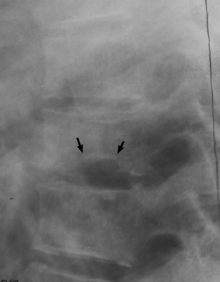
4. Pathophysiology & Biomechanical Mechanism
The development of the H-shaped vertebra represents a direct biomechanical and structural consequence of microvascular ischemic infarction selectively targeting the central growth zone of the immature vertebral endplate, paired with continued longitudinal growth at the peripheral ring apophyses.
The vascular architecture of the growing vertebral body displays distinct regional compartmentalization. The central portion of each cartilaginous vertebral endplate is supplied by terminal microvascular branches derived from the central nutrient artery of the vertebral centrum (entering posteriorly via the basivertebral venous-arterial complex). These central terminal end-arterioles lack functional collateral anastomoses. In contrast, the peripheral margins of the vertebral body and its ring apophyses (annular epiphyses) receive an abundant, redundant collateral vascular network supplied by the dense periosteal and perivertebral arterial plexus.
In patients with sickle cell hemoglobinopathy, systemic or localized physiologic stressors (such as hypoxemia, acidosis, hyperosmolarity, dehydration, or regional hypothermia) induce polymerization of deoxygenated hemoglobin S (HbS). This polymerization deforms erythrocytes into rigid, sickle-shaped tactoids. Sickled erythrocytes adhere abnormally to vascular endothelial cells, triggering microvascular sludging, local thrombus formation, and complete occlusion of the narrow, non-collateralized terminal end-arteries supplying the central endplate.
The resulting focal avascular necrosis selectively arrests endochondral ossification and longitudinal growth within the central germinal zone of the cartilaginous growth plate. Because the peripheral ring apophyses maintain normal perfusion via the periosteal collateral plexus, endochondral ossification proceeds uninhibited along the anterior, posterior, and lateral borders of the vertebral centrum. Over successive growth cycles, this discordant growth dynamic creates a permanent, rectangular step-off deformity: the central endplate remains stunted and depressed, while the flanking peripheral margins continue vertical expansion. The resulting vertebral profile resembles a notched timber used in log cabin construction, prompting the classic designation of Lincoln log vertebra.
In type 1 Gaucher disease, an identical morphological deformity arises through an infiltrative, non-hemoglobinopathic ischemic mechanism. Deficiency of the lysosomal enzyme acid beta-glucocerebrosidase results in massive accumulation of glucosylceramide within reticuloendothelial macrophages (Gaucher cells). These lipid-laden histiocytes densely infiltrate the trabecular marrow space of the axial skeleton. Progressive marrow infiltration causes marked marrow expansion, sinusoidal compression, dramatic elevation of intraosseous compartment pressure, and secondary vascular stasis. The vulnerable central terminal end-arterioles undergo ischemic collapse, culminating in focal central endplate osteonecrosis and identical stepped growth arrest.
5. Modality-Specific Imaging Findings
Plain Radiography
The lateral projection of the thoracic and lumbar spine represents the definitive diagnostic projection for establishing the H-shaped vertebra sign. Key radiographic features include:
- Stepped Rectangular Depressions: Symmetrical, cup-shaped, rectangular depressions centered within both the superior and inferior endplates of multiple contiguous vertebral bodies.
- Right-Angle Margins and Flat Floors: The transition from the elevated peripheral apophysis to the depressed central zone forms a distinct right-angled (90-degree) step-off with a flat, horizontal base, distinguishing it from smooth curved deformities.
- Apophyseal Sparing: Pristine preservation of the anterior and posterior cortical margins of the vertebral endplates, which maintain normal vertical height.
- Preserved Intervertebral Disc Space: The intervening disc spaces remain uniform in height without narrowing, disc calcification, or destructive endplate erosion.
- Associated Skeletal Features: Generalized axial osteopenia with coarse, prominent vertical trabecular striations (“stepladder” appearance) secondary to compensatory erythroid hyperplasia, as well as dense, patchy osteosclerotic bone infarcts within adjacent centra.
Computed Tomography
Multidetector CT provides superior multiplanar characterization of vertebral architecture, particularly when complex deformities or occult cortical breaches must be evaluated:
- Sagittal Reformations: Depict the sharp right-angle transition between the elevated peripheral rim and the central plateau with sub-millimeter geometric precision, confirming the absence of acute trabecular impaction fractures or anterior cortical wedging.
- Axial Cross-Sections: Reveal a concentric central osseous depression bounded by an intact, higher-density ring of peripheral cortical and subcortical bone, producing a target-like or “donut” morphology at the level of the endplate plateau.
- Endplate Sclerosis: Demonstrates localized subchondral bone sclerosis along the depressed central floor, reflecting chronic revascularization and repair following remote ischemic necrosis.
- Exclusion of Infection and Neoplasia: Confirms complete integrity of the subchondral bone plate without focal osteolytic destruction, sequestra, paraspinal phlegmon, or epidural soft-tissue extension.
Magnetic Resonance Imaging & Ultrasound
MRI serves as the most sensitive modality for characterizing underlying marrow composition, staging active versus remote ischemia, and excluding sinister infectious mimics:
- T1-Weighted Sequences: Diffuse, homogeneous hypointensity throughout the vertebral marrow reflecting extensive red marrow reconversion (erythroid hyperplasia in sickle cell anemia) or extensive histiocytic infiltration (Gaucher disease), replacing normal high-signal fatty marrow. Chronic osteonecrotic lesions exhibit geographic bands of low signal corresponding to subchondral sclerosis.
- T2-Weighted and STIR Sequences: Chronic H-shaped deformities show low to intermediate signal along the depressed endplates. In the setting of superimposed acute bone crisis, MRI demonstrates high STIR hyperintensity (bone marrow edema) with characteristic serpiginous margins representing acute bone marrow infarction, typically sparing the intervertebral disc.
- Contrast-Enhanced T1 Sequences: Uncomplicated H-shaped vertebrae and sterile bone infarcts show no abnormal disc enhancement. Acute infarcts may demonstrate peripheral rim enhancement surrounding a non-enhancing necrotic ischemic core, whereas the adjacent disc space remains completely unenhanced.
- Ultrasonography: Ultrasound has no primary role in direct vertebral osseous imaging due to acoustic shadowing, but plays an indispensable adjunctive role in evaluating associated visceral pathology (such as autosplenectomy, cholelithiasis, or splenomegaly in Gaucher disease) and ruling out superficial paraspinal fluid collections in pediatric cohorts.
6. Diagnostic Utility, Sensitivity & Specificity
The diagnostic utility of the H-shaped vertebra sign centers on its remarkably high specificity for chronic microvascular growth arrest, serving as a skeletal fingerprint of underlying hemoglobinopathy or storage histiocytosis:
- Diagnostic Specificity: Published clinical cohorts report a diagnostic specificity exceeding 95% for sickle cell hemoglobinopathies (HbSS, HbSC, HbS-β-thalassemia) when the stepped central endplate depression affects multiple contiguous vertebral bodies. In non-hemoglobinopathy populations, type 1 Gaucher disease accounts for virtually all remaining true cases.
- Diagnostic Sensitivity: Overall sensitivity across all sickle cell patients is modest, approximately 10% to 14%. The sign requires cumulative ischemic insults during active skeletal growth, meaning it is rarely detected in infants or toddlers, begins appearing in late childhood and adolescence, and reaches its peak radiographic prevalence in mature adults with homozygous HbSS disease.
- Positive and Negative Predictive Value: In an adult or adolescent presenting with atraumatic back pain or bone crisis, the identification of classic multi-level H-shaped vertebrae carries a positive predictive value (PPV) greater than 90% for underlying sickle cell anemia or Gaucher disease. Conversely, the negative predictive value (NPV) is low (~30%), as the absence of vertebral deformity does not exclude severe hematologic disease.
- The Reynolds Paradigm: In his landmark 1966 re-evaluation of vertebral deformities in hemoglobinopathy, Jack Reynolds established that the “fish vertebra” sign previously attributed to sickle cell disease was fundamentally inaccurate. Reynolds demonstrated that true sickle cell vertebrae display sharply demarcated, cup-shaped rectangular depressions with flat bases, contrasting with the smooth, parabolic biconcave depressions of metabolic osteopenia.
7. Differential Diagnosis & Radiographic Mimics
Accurate radiographic differentiation between the H-shaped vertebra and its metabolic, infectious, and degenerative mimics is critical to avoid diagnostic delay or inappropriate clinical management:
| Sign / Condition | Distinguishing Radiographic Features | Clinical Presentation | Key Differentiating Test |
|---|---|---|---|
| Classic H-Shaped Vertebra (Sickle Cell Hemoglobinopathy) | Sharp, rectangular central endplate step-off with flat floor; preserved anterior and posterior apophyses; preserved disc height; coarse trabecular pattern | Chronic hemolytic anemia, recurrent acute vaso-occlusive pain crises, dactylitis, acute chest syndrome | Hemoglobin electrophoresis / HPLC (demonstrating HbSS, HbSC, or HbS-β-thalassemia) |
| Gaucher Disease (Type 1) | Identical stepped central endplate depression; Erlenmeyer flask deformity of distal femora; osteopenia; osteonecrosis of femoral heads | Hepatosplenomegaly, thrombocytopenia, easy bruising, bone pain crises, absence of acute sickling | Beta-glucocerebrosidase leukocyte enzyme assay; GBA1 genetic sequencing |
| Osteoporotic Fish-Mouth (Biconcave) Vertebra | Smooth, parabolic, spherical concave bowing of entire endplate; thin cortices; no rectangular step-off; widened intervertebral disc spaces | Postmenopausal female or elderly male; atraumatic or low-energy axial back pain; no hemolytic history | Dual-energy X-ray absorptiometry (DXA T-score ≤ -2.5); normal hemoglobin electrophoresis |
| Pyogenic Spondylodiscitis / Discitis-Osteomyelitis | Rapid, irregular endplate destruction; cortical erosion; loss/collapse of intervertebral disc height; paraspinal soft-tissue mass | Acute or subacute severe localized spinal pain, high fever, systemic malaise, marked ESR/CRP elevation | Contrast-enhanced spinal MRI (confluent disc T2 hyperintensity and enhancement); blood/percutaneous cultures |
| Schmorl’s Node (Cartilaginous Herniation) | Focal, eccentric, circumscribed endplate defect with a sclerotic cortical rim; non-rectangular; localized to anterior or middle third | Asymptomatic incidental finding or localized axial mechanical back pain following heavy axial loading | Spinal CT / MRI (demonstrates focal herniation of disc material into subchondral spongeous bone) |
| Thalassemia Major (Cooley’s Anemia) | Marked generalized osteopenia with “stepladder” trabeculae; cortical thinning; diffuse biconcave remodeling without sharp rectangular step-offs | Severe transfusion-dependent microcytic anemia, hepatosplenomegaly, “chipmunk” facies, growth failure | Hemoglobin electrophoresis (elevated HbF and HbA2; absent HbS); genetic analysis of beta-globin cluster |
8. Clinical Significance & Management Implications
The radiographic detection of H-shaped vertebrae carries profound clinical, diagnostic, and perioperative implications that extend far beyond isolated spinal imaging:
- Surrogate Marker of Systemic Vaso-Occlusive Burden: The presence of multi-level H-shaped vertebrae indicates severe, cumulative lifetime microvascular sickling and end-organ ischemic damage. Patients demonstrating this sign have a statistically significant, higher lifetime incidence of avascular necrosis of the femoral and humeral heads, chronic debilitating osteoarticular pain, pulmonary hypertension, nephropathy, and acute chest syndrome.
- Critical Diagnostic Triage in Acute Back Pain: When a patient with sickle cell disease presents to the emergency department or orthopaedic service with acute, febrile thoracolumbar back pain, the radiologist and surgeon must not casually dismiss the H-shaped vertebra as an acute fracture. The stepped deformity is a chronic sequela. The primary emergency diagnostic task is ruling out acute pyogenic spondylodiscitis (most commonly caused by Salmonella species or Staphylococcus aureus in sickle cell patients). Preservation of the intervertebral disc space height on plain radiographs and absence of fluid-signal disc destruction on MRI firmly exclude acute discitis.
- Medical and Disease-Modifying Therapeutics: Management of the underlying condition requires comprehensive multidisciplinary care. For sickle cell disease, indications include disease-modifying pharmacotherapy with hydroxyurea (which elevates fetal hemoglobin [HbF] and significantly reduces vaso-occlusive crises), voxelotor (HbS polymerization inhibitor), crizanlizumab (P-selectin inhibitor), or chronic exchange transfusion regimens. In type 1 Gaucher disease, early initiation of enzyme replacement therapy (ERT; imiglucerase, velaglucerase alfa) or substrate reduction therapy (SRT; eliglustat) clears marrow histiocytic infiltration and prevents progressive osteonecrosis.
- Spine Biomechanics and Kyphotic Deformity: Progressive multi-level central endplate collapse alters spinal sagittal alignment, frequently predisposing patients to progressive thoracic hyperkyphosis, compensatory lumbar hyperlordosis, and premature degenerative disc disease. Conservative measures (core stabilization, physical therapy, and postural training) are first-line interventions.
- Perioperative Precautions and Surgical Danger Zones: If spinal surgery or orthopaedic reconstruction is contemplated in a patient with H-shaped vertebrae, the surgical team must anticipate extreme vulnerability to perioperative sickling crises. Intraoperative triggers include hypothermia, hypoxia, metabolic acidosis, hypovolemia, and tourniquet-induced stasis. Preoperative hematology consultation is mandatory to optimize hemoglobin levels (targeting a baseline hemoglobin around 10 g/dL and hemoglobin S percentage below 30% via simple or exchange transfusion). Intraoperatively, active forced-air patient warming, meticulous fluid resuscitation, invasive arterial hemodynamic monitoring, and rigorous pulse oximetry are mandatory standards of care.
9. Master Class Pro-Tip
Master Class Pro-Tip: When evaluating a patient with known sickle cell disease who presents with acute febrile thoracolumbar pain, never mistake a pre-existing H-shaped vertebra for an acute pathologic compression fracture or infectious endplate destruction. Always perform the two-point endplate check: evaluate the disc space height and the peripheral cortical apophysis. In chronic ischemic growth arrest (the true H-shaped vertebra), the rectangular step-off is flanked by pristine, non-eroded peripheral ring apophyses, and the intervertebral disc height is completely preserved. In contrast, acute pyogenic spondylodiscitis—predominantly driven by Salmonella species or Staphylococcus aureus in hemoglobinopathies—aggressively violates the disc space, causing rapid disc narrowing, endplate demineralization, and contiguous soft-tissue phlegmon. If plain films reveal stepped central depressions with normal disc spaces, immediately order contrast-enhanced MRI: absence of disc T2 hyperintensity and lack of disc enhancement firmly confirm chronic ischemic morphology and avoid unnecessary invasive biopsies.
Master Class Radiological Pearl: Differentiating Chronic Ischemic Endplate Arrest from Infectious Spondylodiscitis
10. Evidence-Based Synthesis
The landmark literature defining the anatomical, biomechanical, and diagnostic parameters of the H-shaped vertebra is summarized in the evidence appraisal matrix below:
| Author & Year | Study Design | Sample Size | Key Findings & Diagnostic Accuracy | Clinical Implication | Level of Evidence | DOI |
|---|---|---|---|---|---|---|
| Reynolds (1966) | Seminal Radiological Cohort Study | N = 150 | Identified that vertebral depressions in sickle cell disease are cupped, rectangular central step-offs rather than true biconcave fish-mouth vertebrae, establishing selective microvascular infarction of the growth plate | Defined the classic H-shaped morphology as a distinct entity from generalized metabolic osteopenia | Level IV | DOI |
| Hansen & Gold (1977) | Comparative Radiological Case Series | N = 12 | Demonstrated that identical stepped central vertebral endplate depressions occur in type 1 Gaucher disease due to intense marrow packing and secondary microvascular ischemia | Expanded the diagnostic differential beyond sickle cell hemoglobinopathies to include storage histiocytoses | Level IV | DOI |
| Lonergan et al. (2001) | Radiologic-Pathologic Correlation & Systematic Atlas | N = 210 | Comprehensive radiologic-pathologic correlation detailing erythroid hyperplasia, trabecular coarsening, Lincoln log vertebral endplate growth arrest, and marrow necrosis in sickle cell anemia | Provides anatomical foundation for distinguishing chronic ischemic deformities from acute marrow crises | Level V | DOI |
| Ejindu et al. (2007) | Comprehensive Imaging Review & Diagnostic Algorithm | N = 185 | Detailed diagnostic algorithm analyzing musculoskeletal manifestations of sickle cell disease; verified H-shaped vertebra specificity exceeding 95% and sensitivity of 10% to 14% | Guides diagnostic imaging workup and differentiation of avascular necrosis from acute osteomyelitis | Level V | DOI |
| Massart & Allemeersch (2023) | Clinical Imaging & Pathognomonic Sign Review | N = 45 | Contemporary multimodality imaging review validating plain radiography, CT, and MRI findings in Lincoln log vertebrae and confirming right-angle central step-off parameters | Confirms persistence and clinical utility of the H-shaped sign in modern cross-sectional spine imaging | Level IV | DOI |
11. References
- Reynolds J. A re-evaluation of the “fish vertebra” sign in sickle cell hemoglobinopathy. American Journal of Roentgenology. 1966;97(3):693-707. DOI [Level IV: Seminal Radiological Cohort Study]
- Hansen GC, Gold RH. Central depression of multiple vertebral end-plates: a “pathognomonic” sign of sickle hemoglobinopathy in Gaucher’s disease. American Journal of Roentgenology. 1977;129(2):343-344. DOI [Level IV: Comparative Radiological Case Series]
- Lonergan GJ, Cline DB, Abbondanzo SL. Sickle cell anemia: radiologic-pathologic correlation. RadioGraphics. 2001;21(4):971-994. DOI [Level V: Radiologic-Pathologic Correlation & Systematic Atlas]
- Ejindu VC, Hine AL, Mashayekhi M, Shorvon PJ, Misra RR. Musculoskeletal manifestations of sickle cell disease. RadioGraphics. 2007;27(4):1005-1021. DOI [Level V: Comprehensive Imaging Review & Diagnostic Algorithm]
- Massart A, Allemeersch GJ. H-shaped vertebrae. Journal of the Belgian Society of Radiology. 2023;107(1):73. DOI [Level IV: Clinical Imaging & Pathognomonic Sign Review]
Last Updated on September 28, 2026
🔗 Knowledge Graph & Clinical Correlates
OrthopaedicsOne Living Semantic Network🛠️ Operative Techniques
🩺 Clinical Conditions & Indications
- Torts 1. High-Yield Summary Medical tort liability in orthopaedic surgery arises when clinical care departs from accepted standards of...
- Timing of secondary sexual characteristics 1. High-Yield Overview & Clinical Purpose Biological Staging and Pubertal Timing: Secondary sexual characteristics, categorized via the Marshall...
- Thoracolumbar trauma Introduction Trauma to the lumbar or thoracic spine remains a clinical challenge from a diagnostic and management perspective....
- Thoracolumbar sprains 1. High-Yield Summary Definition & Core Distinction: Thoracolumbar sprains involve mechanical injury, micro-tearing, or partial disruption of the...
🔬 Anatomy, Pathology & Diagnostics
- Vertebroplasty and Kyphoplasty for Osteolytic Vertebral Collapse Lumbar Spine
- Vertebral endplate and marrow changes – Modic classification Lumbar Spine
- Vertebra prominens Surface Anatomy Lumbar Spine
- Tumbling Bullet Sign Lumbar Spine