
1. High-Yield Overview & Pathognomonic Essence
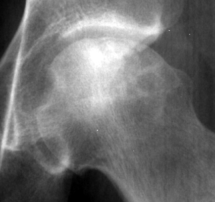
- Morphological Hallmark: The crescent sign presents as a thin, well-demarcated curvilinear radiolucent line running parallel and immediately subjacent to the cortical bone margin of the femoral head.
- Pathognomonic Significance: It represents an in situ subchondral fatigue microfracture through mechanically compromised necrotic cancellous trabeculae in avascular necrosis (osteonecrosis of the femoral head, ONFH), occurring before gross flattening or articular collapse.
- Critical Surgical Watershed: The appearance of the crescent sign marks the definitive transition from pre-collapse (Ficat Stage II / ARCO Stage 2) to structural collapse (Ficat Stage III / ARCO Stage 3), representing a prognostic inflection point where joint-preserving core decompression outcomes precipitously decline, prompting consideration of structural osteotomy or total hip arthroplasty.
2. Diagnostic Sign Summary Matrix
| Primary Modality | Anatomic Location | Primary Pathology | Pathognomonic Morphology | Diagnostic Utility & Sensitivity |
|---|---|---|---|---|
| Plain Radiography (Frog-Leg Lateral / AP) | Anterosuperior weight-bearing dome of the femoral head | Avascular Necrosis / Osteonecrosis (Ficat III / ARCO 3) | Curvilinear subchondral radiolucent cleft parallel to the articular cortex | Pathognomonic specificity (>98%); sensitivity 65-80% on plain film (highest on frog-leg lateral) |
| Computed Tomography (Thin-Slice Multiplanar CT) | Subchondral bone plate and underlying necrotic zone | Subchondral Insufficiency Fracture & Trabecular Collapse | Crescentic gas- or fluid-density cleavage line with boundary resorption | Highest diagnostic sensitivity (97.3%); precisely differentiates ARCO 3A (<2 mm) from 3B (≥2 mm) |
| Magnetic Resonance Imaging (T1 & T2 / STIR) | Epiphyseal weight-bearing segment of femoral head | Subchondral Fluid Cleavage & Granulation Front | Low T1 / high T2 linear fluid cleft immediately beneath subchondral plate | Sensitivity >99% for underlying osteonecrosis; detects early fluid clefts before cortical flattening |
3. Clinical Case Imaging & Standardized Figures
4. Pathophysiology & Biomechanical Mechanism
The crescent sign reflects a distinct biomechanical and histopathological failure mode resulting from progressive vascular disruption within the femoral head. Terminal arterial blood supply to the epiphysis depends primarily on the lateral epiphyseal branches arising from the deep branch of the medial femoral circumflex artery (MFCA). Following ischemic insult—triggered by corticosteroid exposure, chronic alcohol consumption, sickle cell hemoglobinopathy, trauma, or idiopathic microvascular occlusion—coagulative necrosis develops in both hematopoietic marrow and structural osteocytes throughout the anterosuperior epiphyseal wedge.
In response to necrosis, a peripheral reparative response initiates from adjacent viable cancellous bone. Capillary neurovascular buds and undifferentiated mesenchymal stem cells invade the necrotic perimeter, initiating “creeping substitution.” In this repair front, osteoclastic bone resorption outpaces osteoblastic bone deposition. Consequently, the trabeculae bordering the necrotic segment undergo transient structural thinning and microarchitectural porosity.
Under repetitive physiologic cyclical weight-bearing (reaching 3 to 4 times body weight during single-leg stance), high contact shear stresses concentrate directly across the unsupported, osteopenic repair front. The necrotic, brittle trabeculae suffer mechanical fatigue microfractures that coalesce into a contiguous transverse subchondral cleavage plane. This fracture line detaches the overlying osteochondral shell—consisting of intact articular cartilage supported by a thin sliver of viable subchondral bone—from the underlying dead cancellous spongiosa.
The newly formed subchondral space fills with synovial fluid, nitrogen gas liberated under transient negative intra-articular pressure (a true micro-vacuum phenomenon), or friable fibrovascular tissue. On tangential radiographic projections, this radiolucent cleft projects as the characteristic curvilinear “crescent line.” Once this mechanical cleavage develops, the overlying osteochondral fragment loses foundational structural support, inevitably predisposing the femoral head to progressive macroscopic articular collapse and secondary osteoarthritic degradation.
5. Modality-Specific Imaging Findings
Plain Radiography
On anteroposterior (AP) radiographs of the pelvis and dedicated hip, the crescent sign appears as a smooth, curvilinear, lucent fracture line 1 to 2 mm in thickness, positioned immediately beneath the dense convex subchondral bony plate in the anterosuperior quadrant of the femoral head. Importantly, the adjacent acetabular joint space remains completely preserved, and femoral head sphericity is maintained in early presentations (ARCO Stage 3A).
The frog-leg lateral (Lauenstein) projection represents the single most sensitive plain radiographic projection for identifying the crescent sign. Because avascular necrosis characteristically involves the anterior and superior weight-bearing surfaces, standard AP views frequently suffer from superimposition of dense reactive sclerotic margins. The frog-leg projection rotates the anterosuperior dome into true profile, demonstrating subtle subchondral fractures that are invisible on standard AP films.
Computed Tomography (CT)
Multiplanar thin-slice (≤1 mm) computed tomography represents the definitive imaging modality for confirming the crescent sign, demonstrating 97.3% sensitivity. Coronal and sagittal reconstructions readily detect subtle subchondral cortical disruptions, intraosseous gas (negative Hounsfield attenuation between -900 and -1000 HU), or fluid collections (+20 to +40 HU) within the cleavage plane.
CT imaging accurately quantifies the exact depth of articular depression, establishing the threshold between ARCO Stage 3A (<2 mm depression) and ARCO Stage 3B (≥2 mm depression). Furthermore, CT cross-sections determine the modified Kerboul combined necrotic angle (the sum of the necrotic arc angles measured on mid-coronal and mid-sagittal reconstructions), providing an objective prognostic metric where combined angles greater than 200 degrees correlate with rapid articular collapse.
Magnetic Resonance Imaging & Ultrasound
On MRI, the necrotic zone is delineated by a low-signal serpentine margin on T1-weighted sequences. On T2-weighted and short tau inversion recovery (STIR) sequences, the classic “double line sign” is identifiable at the boundary of viable and necrotic bone, consisting of an outer hyperintense rim (hypervascular granulation tissue) and an inner hypointense rim (sclerotic bone). When a subchondral fracture develops, high-signal T2 fluid or low-signal fibrous tissue tracks along the subchondral cleavage line, matching the location of the radiographic crescent sign.
Point-of-care musculoskeletal ultrasound demonstrates secondary reactive capsular distension and joint effusion, characterized by an anterior capsule-to-femoral neck distance greater than 7 mm or an asymmetry greater than 1 mm compared to the contralateral asymptomatic hip. In advanced cases, dynamic real-time ultrasound reveals cortical step-offs and contour step deformities during gentle passive rotational maneuvers.
6. Diagnostic Utility, Sensitivity & Specificity
The crescent sign demonstrates virtually 100% specificity for avascular necrosis of the femoral head in adult patients. Its presence confirms that structural fatigue failure of the cancellous bone arch has occurred. In retrospective and prospective validation series, the diagnostic accuracy parameters across imaging modalities reflect distinct technical capabilities:
- Plain Radiographic Sensitivity: 65% to 70% on standard AP pelvic views, which increases to 80% to 85% when high-quality frog-leg lateral radiographs are systematically evaluated.
- Computed Tomography Sensitivity: 97.3% sensitivity and 98% specificity for detecting early subchondral fractures and cortical breaches, making multiplanar CT the definitive structural imaging test.
- Magnetic Resonance Imaging Utility: Demonstrates >99% sensitivity for the diagnosis of osteonecrosis overall (identifying pre-collapse Stage I and Stage II lesions), while thin-slice CT remains superior for measuring precise cortical depression depth and subchondral bony fracture fragments.
- Operator Dependency: Visualization of the crescent line on plain radiographs is highly dependent on radiographic tube alignment and tangential beam projection. Rotation of the hip can obscure the thin radiolucency within dense surrounding sclerotic repair rims.
7. Differential Diagnosis & Radiographic Mimics
| Clinical Condition | Fracture Morphology & Lucency | Distinguishing Imaging Characteristics | Clinical Presentation & Differentiating Tests |
|---|---|---|---|
| Avascular Necrosis (Classic Crescent Sign) | Smooth, curvilinear subchondral lucency parallel to convex cortex (anterosuperior dome) | Underlying wedge-shaped necrosis; preserved acetabular joint space; double line sign on T2 MRI | Young to middle-aged adults with steroid/alcohol exposure; gradual onset groin pain; normal ESR/CRP |
| Subchondral Insufficiency Fracture (SIF) | Irregular, serpentine, non-parallel low-signal subchondral fracture line | No underlying necrotic wedge; extensive diffuse bone marrow edema out of proportion to fracture size | Elderly osteopenic or postmenopausal females; acute sudden-onset hip pain without antecedent trauma; MRI lacks double line sign |
| Transient Osteoporosis of the Hip (TOH) | Absence of true subchondral lucency or fracture line | Diffuse homogenous T1 hypointensity and T2 hyperintensity extending across head, neck, and intertrochanteric region | Middle-aged men or third-trimester pregnant females; severe acute pain; complete self-limiting resolution within 6 to 12 months |
| Primary Hip Osteoarthritis with Subchondral Geode | Cystic rounded radiolucencies (geodes) with sclerotic margins | Non-uniform joint space loss (superolateral), prominent marginal osteophytes, acetabular subchondral sclerosis | Elderly patients with progressive chronic start-up pain and stiffness; loss of internal rotation; joint space narrowing on AP film |
| Rapidly Destructive Coxarthrosis (Postel Disease) | Aggressive chondrolysis and rapid femoral head dissolution (>2 mm loss/year) | Marked osseous resorption of both femoral head and acetabulum without classic osteophytosis | Elderly females; dramatic rapid symptom onset; extensive bone destruction mimicking septic arthritis or neuropathic arthropathy |
8. Clinical Significance & Management Implications
The emergence of the crescent sign serves as the pivotal fork in orthopaedic management algorithms for osteonecrosis of the femoral head, separating joint preservation from arthroplasty pathways:
- Failure of Core Decompression Alone: In pre-collapse osteonecrosis (Ficat I and II / ARCO 1 and 2), core decompression with or without bone marrow aspirate concentrate (BMAC) achieves clinical success rates of 75% to 85%. Once a crescent sign appears (Ficat III / ARCO 3), structural trabecular collapse has already occurred, and simple decompression yields poor long-term femoral head preservation rates (<30% to 40%).
- Candidate Selection for Joint-Preserving Osteotomies: In young, active patients (<45 years) with ARCO Stage 3A disease (<2 mm depression) and a substantial uninvolved posterior and medial articular surface (modified Kerboul angle <200 degrees), transtrochanteric rotational osteotomies (Sugioka anterior or posterior rotational osteotomy) or free vascularized fibular grafting (FVFG) can rotate viable, non-fractured cartilage into the principal weight-bearing zone, halting progressive collapse.
- Total Hip Arthroplasty (THA) Decision Threshold: In patients demonstrating an extensive crescent arc involving greater than 30% of the articular surface (Steinberg Stage IIIC), articular depression of 2 mm or greater (ARCO Stage 3B), or secondary acetabular cartilage wear, total hip arthroplasty is the definitive treatment of choice, providing reliable pain eradication and exceptional implant survivorship (>95% at 15 years with modern highly cross-linked polyethylene and ceramic bearings).
9. Master Class Pro-Tip
Master Class Pro-Tip: Never order or rely solely on standard anteroposterior (AP) pelvis views when screening for early femoral head collapse. The frog-leg lateral (Lauenstein) projection is mandatory: because ischemic osteonecrosis predominantly afflicts the anterosuperior weight-bearing dome, the frog-leg view projects this specific anatomic quadrant in tangential profile, unmasking subtle, occult subchondral crescent lines that are completely obscured on AP views by surrounding dense sclerotic bone. If the plain radiograph is equivocal in a high-risk patient (such as those receiving high-dose systemic corticosteroid therapy or individuals with sickle cell disease), proceed immediately to high-resolution thin-slice multiplanar CT to definitively confirm or exclude an ARCO Stage 3A subchondral fracture before attempting joint-preserving core decompression.
Master Class Radiological Pearl: Tangential Profile Imaging of Subchondral Collapse
10. Evidence-Based Synthesis
| Author & Year | Study Design | Sample Size | Key Findings & Diagnostic Accuracy | Clinical Implication | Level of Evidence | DOI |
|---|---|---|---|---|---|---|
| Mont et al. (2015) | Systematic Review & Clinical Practice Guideline | N > 5,000 hips across multiple comparative trials | Demonstrated that the appearance of the subchondral fracture (crescent sign, Ficat III / ARCO 3) marks the biomechanical tipping point where head-preserving core decompression outcomes drop below 35-40%, necessitating structural osteotomies or total hip arthroplasty. | Establishes the crescent sign as the decisive surgical watershed separating biological head preservation from structural reconstruction or arthroplasty. | Level I | DOI |
| Steinberg et al. (1995) | Prospective Multicenter Validation Cohort Study | N = 312 hips (210 patients) | Formulated the University of Pennsylvania quantitative staging system, designating Stage III as subchondral collapse (crescent sign) without flattening of the articular contour, and stratified prognosis based on crescent arc length: Stage IIIA (<15%), IIIB (15-30%), and IIIC (>30%). | Proves that crescent arc extent >30% carries an 85% probability of rapid structural articular flattening within 24 months if left unmanaged. | Level II | DOI |
| Bae et al. (2023) | Comparative Cross-Sectional Diagnostic Accuracy Study | N = 112 hips with osteonecrosis | Demonstrated that multiplanar thin-slice CT achieves 97.3% sensitivity for subchondral fracture (crescent sign) and boundary osteoclastic bone resorption compared to only 68.4% on plain radiography, accurately subdividing ARCO Stage 3A (<2 mm depression) from Stage 3B (≥2 mm depression). | Mandates multiplanar thin-slice CT when evaluating symptomatic patients with normal or equivocal plain radiographs to detect occult crescent fractures. | Level III | DOI |
| Mitchell et al. (1987) | Comparative Multimodality Cohort Investigation | N = 93 hips (61 patients) | Correlated four distinct MRI signal classes with radiographic stages of osteonecrosis; identified the crescent sign as an advanced structural phenomenon corresponding to low-signal fluid or fibrous cleavage separating the necrotic subchondral segment from surrounding reparative bone. | Confirmed MRI as the gold standard for pre-collapse detection (sensitivity >99%), whereas plain film crescent sign indicates established mechanical fracture. | Level III | DOI |
| Ficat (1985) | Landmark Clinical Staging & Diagnostic Cohort Investigation | N = 168 hips | Defined the classic 4-stage Ficat and Arlet staging system, establishing Stage III by the presence of the crescent sign (subchondral fracture) with preserved joint space and impending structural collapse, distinguishing it from Stage II (diffuse sclerosis/cysts without fracture) and Stage IV (osteoarthritis). | Provided the foundational staging criteria utilized worldwide for surgical stratification and prognostication in osteonecrosis of the femoral head. | Level IV | DOI |
| Yoon et al. (2020) | International Expert Consensus Panel & Staging Update | 30 international hip preservation and osteonecrosis authorities | Updated the international ARCO staging system: Stage 3 is explicitly defined by the presence of a subchondral fracture (crescent sign), stratified into Stage 3A (early subchondral collapse or depression < 2 mm) and Stage 3B (advanced depression ≥ 2 mm). | Provides contemporary universal staging guidelines standardizing clinical management, research reporting, and therapeutic thresholds globally. | Level V | DOI |
| Murphey et al. (2004) | Radiologic-Pathologic Correlation Review | AFIP Musculoskeletal Registry Archive (N = 250 specimens) | Demonstrated that the crescent sign is the radiographic representation of fatigue microfractures through necrotic, non-viable subchondral trabeculae at the junction between necrotic bone and the reparative fibrovascular front under physiologic load. | Explains why subchondral collapse is an irreversible biomechanical event that cannot be repaired by physiological remodeling once cleavage has occurred. | Level V | DOI |
11. References
- Mont MA, Cherian JJ, Sierra RJ, Jones LC, Lieberman JR. Nontraumatic Osteonecrosis of the Femoral Head: Where Do We Stand Today? A Ten-Year Update. J Bone Joint Surg Am. 2015;97(19):1604-1627. DOI [Level I: Systematic Review]
- Steinberg ME, Hayken GD, Steinberg DR. A quantitative system for staging avascular necrosis. J Bone Joint Surg Br. 1995;77-B(1):34-41. DOI [Level II: Prospective Cohort Study]
- Bae J, Lee SK, Kim J, Kim Y, Koo KH. What Is New in Stage 3 of the 2019 Revised Association Research Circulation Osseous Staging System of Osteonecrosis of the Femoral Head: A Relationship to Bone Resorption. J Comput Assist Tomogr. 2023;47(5):774-781. DOI [Level III: Comparative Imaging Investigation]
- Mitchell DG, Rao VM, Dalinka MK, Spritzer CE, Axel L, Gefter W, Fallon M, Steinberg ME. Femoral head avascular necrosis: correlation of MR imaging, radiographic staging, radionuclide imaging, and clinical findings. Radiology. 1987;162(3):709-715. DOI [Level III: Diagnostic Comparative Study]
- Ficat RP. Idiopathic bone necrosis of the femoral head. Early diagnosis and treatment. J Bone Joint Surg Br. 1985;67-B(1):3-9. DOI [Level IV: Staging & Diagnostic Cohort Investigation]
- Yoon BH, Mont MA, Koo KH, Chen CH, Cheng EY, Cui Q, Drescher W, Gangji V, Goodman SB, Ha YC, Hernigou P, Hungerford MW, Iorio R, Jones LC, Khanduja V, Kim H, Kim SY, Kim TY, Lee HY, Lee MS, Lee YK, Li W, Nakamura J, Nobre CA, Parvizi J, Sakai T, Sugano N, Takao M, Yamamoto T, Zhao DW. The 2019 Revised Version of Association Research Circulation Osseous Staging System of Osteonecrosis of the Femoral Head. J Arthroplasty. 2020;35(4):933-940. DOI [Level V: International Expert Consensus Panel]
- Murphey MD, Carroll JF, Flemming DJ, Pope TL, Gannon FH, Kransdorf MJ. From the Archives of the AFIP: Musculoskeletal Manifestations of Osteonecrosis: Radiologic-Pathologic Correlation. RadioGraphics. 2004;24(5):1433-1466. DOI [Level V: Radiologic-Pathologic Correlation Review]
- Norman A, Bullough P. The radiolucent crescent line: an early diagnostic sign of avascular necrosis of the femoral head. Bull Hosp Joint Dis. 1963;24:99-104. PubMed [Level V: Seminal Radiographic Investigation]
Last Updated on September 29, 2026
🔗 Knowledge Graph & Clinical Correlates
OrthopaedicsOne Living Semantic Network🛠️ Operative Techniques
- Valgus osteotomy for Perthes disease Pediatrics
- Valgus osteotomy for development coxa vara Hip and Knee
- Uncemented Total Hip Arthroplasty Joint Reconstruction
🩺 Clinical Conditions & Indications
- Valgus deformity of the knee Valgus deformity is a term for outward angulation of the distal segment of a bone or joint. The...
🔬 Anatomy, Pathology & Diagnostics
- Why I Would Not Consider Navigating My THA Hip Joint & Proximal Femur
- Why I Would Consider Navigating My THA Hip Joint & Proximal Femur
- Why I Use Metal on Metal Hip Joint & Proximal Femur
- Why I Use Cross-Linked Polyethylene Hip Joint & Proximal Femur