
Although not common, revision procedures in paediatric scoliosis surgery present a significant challenge to the treating surgeons. This article reviews the common indications for complex spinal reconstructions and revisions, outlines an approach to the deformities, and describes the available osteotomies and their indications to help achieve the reconstruction goals.
Normal Alignment
Understanding the normal spinal alignment will help with surgical planning. In the sagittal plane, the thoracolumbar junction T10 to L2 is neutral. There is approximately 20 to 30° more lumbar lordosis than thoracic kyphosis. A balanced spine is most often achieved when pelvic incidence + thoracic kyphosis (T5-T12) – lumbar lordosis (T12-S1) ? 45° (PI + TK – LL ? 45°). The spine is straight in the coronal plane, with the shoulders and pelvis level. The C7 plumb line should bisect the sacrum in both planes.
Indications
The common indications for revision procedures and spinal osteotomies include the following:
- Coronal and/or sagittal decompensation, generally secondary to previous fusions that stopped short of the stable vertebra or were underecontoured in the sagittal plane
- Curves fused at a young age (prior to closure of the triradiate cartilage) that continue to “crankshaft,” often despite anterior and posterior fusions
- Infected cases or cases with neurological changes where desired corrections were lost in order to eradicate infection or maintain spinal cord function
- Cases with loss of fixation and subsequent loss of correction
Deformity Considerations
- Location of the apex of the deformity in the sagittal and coronal planes
- Magnitude of the deformity
- Flexible or rigid?
- Global or focal?
- Sagittal, coronal, or both?
- Coronal balance: Are the shoulders and pelvis level; is there coronal translation?
- Sagittal balance: Does the C7 plumb line bisect the L5-S1 disc?
- Compensated or non-compensated plumb line: Are mobile regions of the spine compensating?
- Compensation possible: Can the mobile regions of the spine contribute to the deformity correction?
Osteotomy Options
In the presence of rigid kyphosis, correction should include either lengthening of the anterior column, shortening of the posterior column, or a combination of the two. Similarly, rigid coronal plane deformities will require concave lengthening, convex shortening, or both. The differential between the lengthening and the shortening will provide the angular correction. The goal will be to utilize the available osteotomies to achieve the desired correction.
Types of Osteotomies
Smith-Petersen Osteotomy (SPO, termed a Ponte if performed through non-fused segments)
- Posterior shortening through resection of the facet joints
- Lengthening anteriorly occurs through the disc space, which must be mobile to achieve sagittal plane correction
- Middle column acts as the fulcrum
- Multiple levels often required to achieve adequate correction
- Expected correction: Sagittal and coronal plane, 5 – 10° per level
Pedicle Subtraction Osteotomy (PSO)
- Posterior closing wedge osteotomy achieved through resection of the posterior elements, complete resection of the pedicles, and triangular resection through the vertebral body
- Can be asymmetric to provide convex closure and coronal plane correction
- Anterior column remains intact and acts as a hinge in osteotomy closure
- Pedicles resected to allow one foramen to accommodate two nerve roots
- Expected correction: Sagittal plane, 25 – 35°; coronal plane, < 15°
Vertebral column resection (VCR)
- Resection of a complete vertebra to allow multi-planar correction
- Preferred approach is through an all-posterior (costaotransversectomy) approach
- Interbody device (cage) inserted to direct correction
- If primarily sagittal plane desired, cage placed anteriorly
- If primarily coronal plane desired, cage placed in posterior concavity
- Expected correction: Sagittal plane, 30 – 50°; coronal plane, < 30°
Case Examples
Case 1
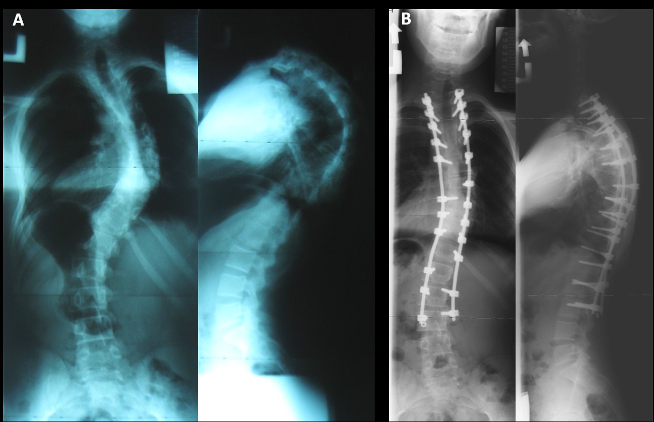
A 17-year-old boy was treated with T1-T7 laminectomy and myelotomy for an astrocytoma at age 18 months. He developed a progressive kyphoscoliosis over the ensuing years with non-debilitating thoracic pain. Radiographs show a 90° mid-thoracic kyphosis with a 70° right thoracic scoliosis, with minimal correction on a bolster supine hyperextension view and on standing side benders. Hyperextension of the lumbar discs compensates for global sagittal balance.
The goal in this case is to improve the thoracic kyphosis and the scoliosis by approximately 50%. Without previous fusions, perhaps up to 20° will be correctable through positioning and instrumentation. This would leave about 20 to 30° of deformity to correct through osteotomies.
This deformity could be addressed either through multiple SPOs with convex closure, an offset PSO to provide both sagittal and coronal correction, or a VCR, if intra-operatively the deformity is felt to be too stiff to allow any passive correction.
With a previous laminectomy extending from T1-T7, it was decided to proceed with an offset T7 PSO. The instrumentation was carried out from T2 to L3. L3 was felt to be the stable vertebra in both planes. The sagittal and coronal corrections are balanced. The preoperative lumbar hyperextension has normalized (Figures 2a-b).

Figures 2a-b. Preoperative (A) and postoperative (B) standing AP and lateral radiographs of a post-laminectomy kypho-scolisosis following offset PSO.
Case 2
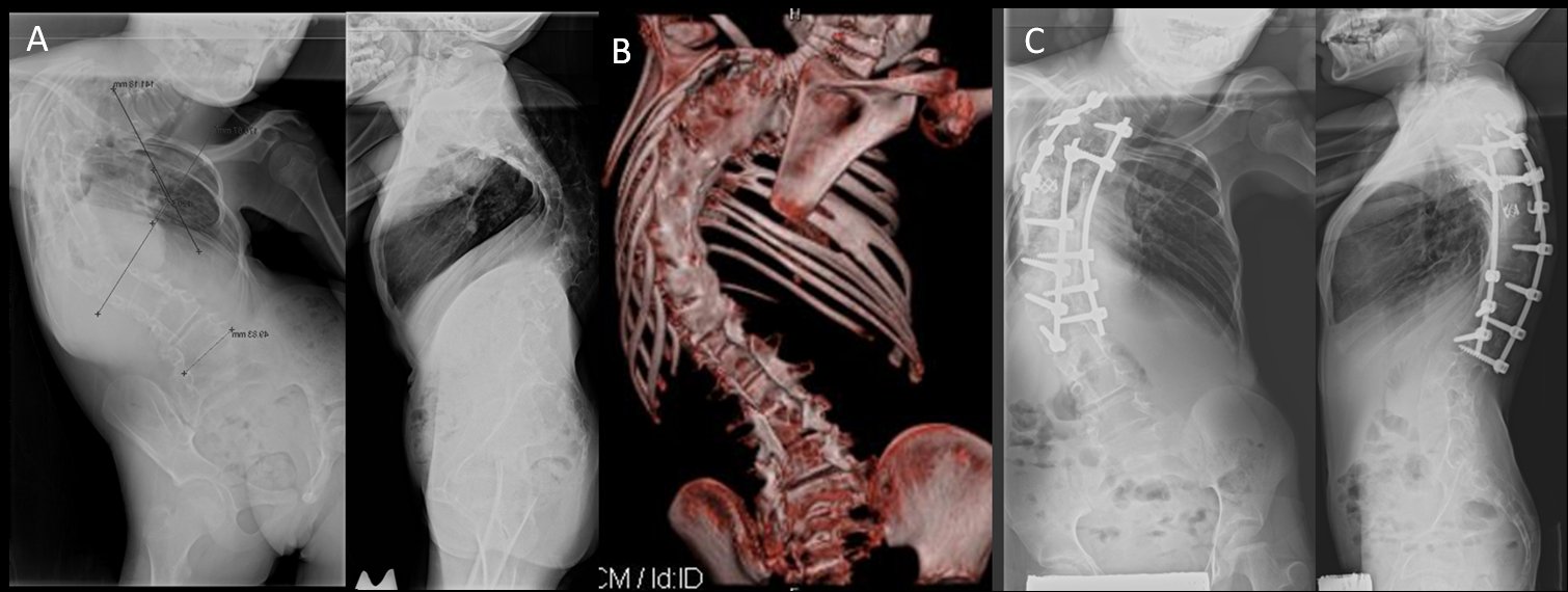
An 11-year-old girl with a history diastomelia and tethered cord developed severe crankshafting and coronal imbalance following previous anterior and posterior spinal fusions for severe progressive scoliosis at age 6. The goal of this case was primarily to normalize the severe coronal imbalance.
Expecting approximately 40° per VCR, two osteotomies were performed, staged at 6-month intervals. Posterior cage placement in the concavity maximized coronal correction (Figures 3a-c).
Figures 3a-c. Preoperative (A), 3D coronal reconstruction (B), and postoperative views (C) following two VCRs performed at six month intervals. Note more normalized sagittal and coronal balance.
Conclusions
Rigid coronal and sagittal plane deformities occur with congenital, previously fused, and inherently stiff curves. These cases present a particular challenge as the usual manoeuvres of derotation, translation, in situ bending, traction, and even anterior release will not provide any significant correction. A strong understanding of the normal spinal alignment and the available osteotomies will help lead to a well thought out plan to provide the patient with a safely corrected and balanced spine.
References
Bernhardt M., Bridwell K.H. Segmental Analysis of the Sagittal Plane Alignment of the Normal Thoracic and Lumbar Spines and Thoracolumbar Junction. Spine 1989: 14:717-721.
Booth K.C., Bridwell K.H., Lenke L.G., et al. Complications and predictive factors for the successful treatment of flatback deformity (fixed sagittal imbalance). Spine 1999;24:1712–20.
Bradford D.S., Boachie-Adjei O. One-stage anterior and posterior hemivertebral resection and arthrodesis for congenital scoliosis. J Bone Joint Surg Am 1990;72:536–40.
Bridwell. K.H. Decision Making Regarding Smith-Petersen vs. Pedicle Subtraction Osteotomy vs. Vertebral Column Resection for Spinal Deformity. Spine2006; 31:S171–S178
Bridwell K., Lewis S.J., Edwards C., et al. Complications and outcomes of pedicle subtraction osteotomies for fixed sagittal imbalance. Spine 2003;28: 2093–101.
Bridwell K.H., Lewis S.J., Lenke L.G., et al. Pedicle subtraction osteotomy for the treatment of fixed sagittal imbalance. J Bone Joint Surg Am 2003;85:454–63.
Bridwell K.H., Lewis S.J., Rinella A., et al. Pedicle subtraction osteotomy for the treatment of fixed sagittal imbalance. Surgical technique. J Bone Joint Surg Am 2004;86–A(suppl 1):44 –50.
Cho K., Bridwell K.H., Lenke L.G., et al. Comparison of Smith-Petersen versus pedicle subtraction osteotomy the correction of fixed sagittal imbalance. Spine 2005;30:2030–7.
Kim Y J., Bridwell K.H., Lenke L.G., Rhim S., Cheh G. An Analysis of Sagittal Spinal Alignment Following Long Adult Lumbar Instrumentation and Fusion to L5 or S1: Can We Predict Ideal Lumbar Lordosis? Spine 2006;31:2343–2352
Rose P.S., Bridwell K.H., Lenke L.G., Cronen G.A., Mulconrey D.S., Buchowski J.M., Kim Y.J. Role of pelvic incidence, thoracic kyphosis, and patient factors on sagittal plane correction following pedicle subtraction osteotomy. Spine. 2009;34:785-79
Smith-Petersen M.N., Larson C.B., Aufranc O.E. Osteotomy of the spine for correction of flexion deformity in rheumatoid arthritis. J Bone Joint Surg Am 1945;27:1–11.
Suk S.I., Kim J.H., Kim W.J., et al. Posterior vertebral column resection for severe spinal deformities. Spine 2002;27:2374–82.
Reprinted with permission from the Summer 2009 issue of COA Bulletin
Attachments:

Last Updated on June 27, 2024
🔗 Knowledge Graph & Clinical Correlates
OrthopaedicsOne Living Semantic Network🛠️ Operative Techniques
🩺 Clinical Conditions & Indications
- Torts 1. High-Yield Summary Medical tort liability in orthopaedic surgery arises when clinical care departs from accepted standards of...
- Timing of secondary sexual characteristics 1. High-Yield Overview & Clinical Purpose Biological Staging and Pubertal Timing: Secondary sexual characteristics, categorized via the Marshall...
- Thoracolumbar trauma Introduction Trauma to the lumbar or thoracic spine remains a clinical challenge from a diagnostic and management perspective....
- Thoracolumbar sprains 1. High-Yield Summary Definition & Core Distinction: Thoracolumbar sprains involve mechanical injury, micro-tearing, or partial disruption of the...
🔬 Anatomy, Pathology & Diagnostics
- Vertebroplasty and Kyphoplasty for Osteolytic Vertebral Collapse Lumbar Spine
- Vertebral endplate and marrow changes – Modic classification Lumbar Spine
- Vertebra prominens Surface Anatomy Lumbar Spine
- Tumbling Bullet Sign Lumbar Spine