
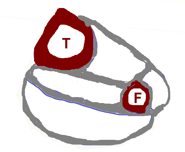
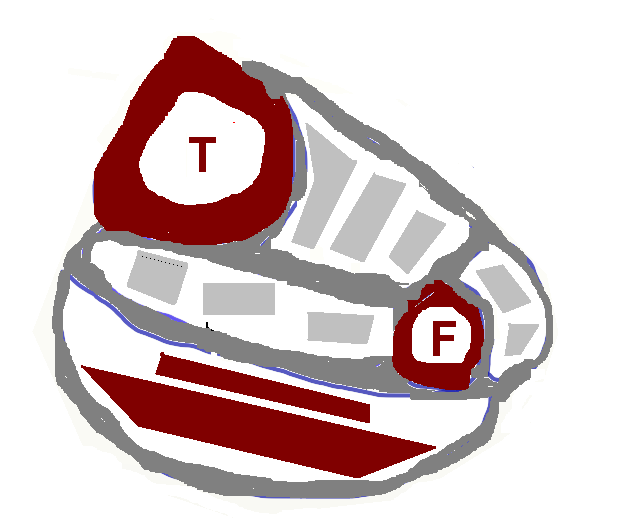
The leg, as seen on an axial cut, is divided into 4 distinct fascial compartments.
 |
| Figure 1. The four compartments of the leg. A line from the tibia to the fibula splits the leg into anterior and posterior. Lateral to the fibula is the lateral compartment. The posterior is further divided into deep and superficial spaces |
 | Attention future surgeons The figures are cartoons, to illustrate the concept of compartments and their contents. The precise layout of the contents, ie. where the muscle originates or where the neurovascular bundle lies, is elided here in the name of simplicity. |
These fascial compartments are separated by septae, tough bands of connective tissue. The muscles within a given fascial compartment usually have similar function and are supplied by the same nerve.
The four compartments of the leg are:
- Anterior
- Lateral
- Deep posterior
- Superficial posterior
| |
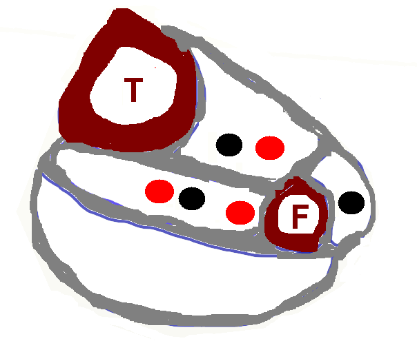
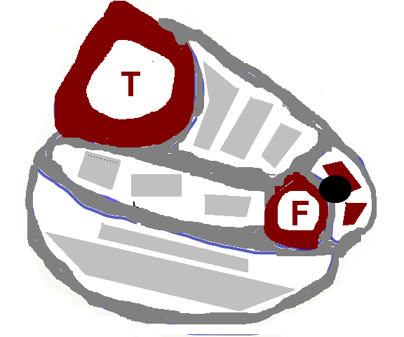
| Figure 2. The “counterintuitive” assortment of blood vessels and arteries. The deep posterior has TWO arteries (the posterior tibial and the peroneal); the lateral has none (but feed off the peroneal). The anterior has the anterior tibial |
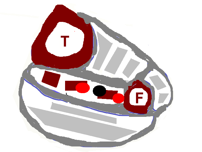
| Attention (NBME) test takers There is no artery or motor nerve in the in the superficial posterior compartment; and the the artery “belonging” to the lateral compartment actually resides in the deep posterior. |
Anterior compartment
| |
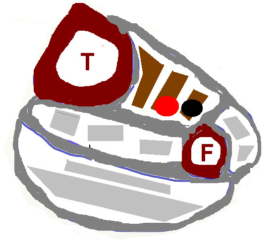
| Figure 3. The anterior compartment,housing the anterior tibial artery, the deep peroneal nerve and 3 muscles: Tibialis anterior, Extensor hallucis longus, Extensor digitorum longus |
The anterior compartment contains the anterior tibial artery, the deep peroneal nerve and the three extensor muscles: tibialis anterior, extensor hallucis longus and extensor digitorum longus. (It also contains the peroneus tertius, which acts like the other peronei (everters), but is supplied by the deep peroneal nerve. That’s too confusing so it’s omitted here. But this factoid will show up on a test, so it’s not really omitted. In fact, this parenthetic aside is longer than the paragraph.)
Lateral compartment
| |
Deep posterior compartment
| |
Superficial posterior compartment
| |
Attachments:

compartments10.PNG (image/png)
compartments3.PNG (image/png)
compartments6.PNG (image/png)
compartments7.PNG (image/png)
compartments8.PNG (image/png)
compartments9.PNG (image/png)
Last Updated on June 27, 2024
🔗 Knowledge Graph & Clinical Correlates
OrthopaedicsOne Living Semantic Network🩺 Clinical Conditions & Indications
- Wigns There are three categories of diagnostic data collected during the physical examination (sign, symptom, and that in between)...
- Why have a privacy policy for my office Original Clinical Contribution: Authored for the OrthopaedicsOne General Orthopaedics Specialty Section. Preserved, digitized, and updated to modern evidence-based...
- Why have a practice website Original Clinical Contribution: Authored for the OrthopaedicsOne General Orthopaedics Specialty Section. Preserved, digitized, and updated to modern evidence-based...
- Who should undergo biopsy of a bone lesion Patient selection for a biopsy is important: It is intended to confirm the radiologic diagnosis in patients with...
💼 Practice Management & Professional Operations
- Winthrop M. Phelps History of Orthopaedics
- Willis Cohoon Campbell History of Orthopaedics
- William Waugh History of Orthopaedics
- William Ward Plummer History of Orthopaedics
- William Thomas Gordon Pugh History of Orthopaedics
- William Stewart Halsted History of Orthopaedics
- William Stevenson Baer History of Orthopaedics
- William Morant Baker History of Orthopaedics