
High-Yield Overview & Pathognomonic Essence
- Pathognomonic Deformity Configuration: Boutonniere (buttonhole) deformity represents a cardinal biomechanical disruption of the digital extensor apparatus characterized by the classic postural triad of proximal interphalangeal (PIP) joint flexion coupled with reciprocal distal interphalangeal (DIP) joint hyperextension and terminal extensor tendon tightness.
- Dual Pathological Mechanisms: The deformity originates through two distinct clinical mechanisms: acute mechanical trauma (extensor Zone III disruption secondary to forced PIP flexion against active extension, dorsal crush lacerations, or volar PIP joint dislocations) and chronic inflammatory arthropathies, predominantly rheumatoid arthritis (RA), wherein proliferative synovial pannus within the retro-extensor pouch stretches, attenuates, and erodes the dorsal apparatus from within.
- Essential Biomechanical Cascade & Management Implication: True boutonniere deformity requires an obligate two-component failure: disruption or attenuation of the central slip insertion into the middle phalanx dorsal tubercle combined with stretching or disruption of the dorsal triangular ligament. This allows the lateral bands to subluxate volarly below the coronal flexion-extension axis of the PIP joint, converting them into paradoxical flexors of the PIP joint while transmitting excessive extensor excursion distally to hyperextend the DIP joint. Early clinical detection via the Elson test mandates 6 weeks of continuous PIP extension splinting with unrestricted active DIP flexion; missed diagnosis permits secondary contractures of the transverse retinacular ligaments and Landsmeer’s oblique retinacular ligaments, converting a flexible deformity into a rigid joint contracture requiring complex surgical reconstruction or arthrodesis.
Diagnostic Sign Summary Matrix
| Primary Modality | Anatomic Location | Primary Pathology | Pathognomonic Morphology | Diagnostic Utility & Sensitivity |
|---|---|---|---|---|
| Clinical Examination (Elson Test) | Proximal and Distal Interphalangeal Joints (Zones III & IV) | Isolated acute disruption or attenuation of the extensor central slip prior to fixed lateral band subluxation | With the PIP joint flexed 90 degrees over a table edge, active PIP extension is weak or absent while the DIP joint exhibits rigid, forceful hyperextension against resistance | Sensitivity ~100%, Specificity ~100% in acute settings; gold standard physical exam test detecting central slip tears before resting deformity or volar lateral band migration occurs |
| High-Resolution Ultrasound (12–18 MHz) | Dorsal Proximal Interphalangeal Joint (Extensor Zone III) | Central slip rupture, dorsal triangular ligament tear, and volar subluxation of lateral bands | Focal hypoechoic or anechoic discontinuity of the central slip over the dorsal PIP joint line; dynamic volar descent of lateral bands beneath the bicondylar rotation axis during flexion; synovial pannus vascularity | Sensitivity 93%, Specificity 100%, Accuracy 95%; exceptional non-invasive dynamic imaging modality confirming occult or equivocal tears in acutely swollen digits |
| Plain Radiography | Digits & Hand (Posteroanterior, True Lateral, Oblique Projections) | Extensor central slip avulsion fracture, inflammatory synovial erosion, and fixed joint contracture | True lateral shows PIP flexion angle with compensatory DIP hyperextension; dorsal avulsion fleck from the base of the middle phalanx; periarticular osteopenia, joint space narrowing, and marginal erosions in rheumatoid arthritis | Sensitivity 100% for bony avulsion fractures and articular destruction; low sensitivity (<25%) for acute isolated soft-tissue tears; primary tool for Burton/Nalebuff staging and joint congruity evaluation |
| Magnetic Resonance Imaging (MRI) | Extensor Hood Apparatus (Zone III to Zone I) | Central slip tendon tear, triangular ligament attenuation, retro-extensor synovial pannus, and chondral loss | T2/PDFS hyperintense fluid signal or complete gap within the central slip insertion at the middle phalanx dorsal base; axial images demonstrate attenuation of triangular ligament and volar migration of lateral bands; dorsal synovial pannus enhancement | High multiplanar contrast resolution; accurately details soft-tissue tendon retraction, collateral ligament integrity, occult cartilage loss, and flexor/extensor tenosynovitis for complex preoperative planning |
Clinical Case Imaging & Standardized Figures
Radiographic assessment of suspected boutonniere deformity requires dedicated multi-view evaluation of the affected digit, with a true lateral projection of the isolated finger representing the single most critical diagnostic view. In acute traumatic settings, plain radiographs primarily evaluate for cortical avulsion fractures at the dorsal base of the middle phalanx (the bony boutonniere injury) and exclude joint subluxation or dislocation. In chronic inflammatory conditions, such as rheumatoid arthritis, plain radiography characterizes the degree of articular destruction, joint space loss, and periarticular demineralization, establishing the anatomic stage and guiding conservative versus operative decision-making.

Figure 1 illustrates the definitive postural alignment of an established boutonniere deformity on a true lateral radiographic projection. The proximal interphalangeal joint is held in substantial flexion (measuring approximately 45 degrees), while the distal interphalangeal joint exhibits reciprocal hyperextension (approximately 20 degrees). Note the prominent dorsal contour of the proximal phalangeal condyles, which have herniated between the attenuated lateral bands in a classic buttonhole configuration. In addition to the postural malalignment, the PIP articulation demonstrates uniform joint space narrowing, subchondral sclerosis, and subtle cortical erosion at the dorsal enthesis of the middle phalanx, confirming an underlying inflammatory rheumatoid arthropathy.
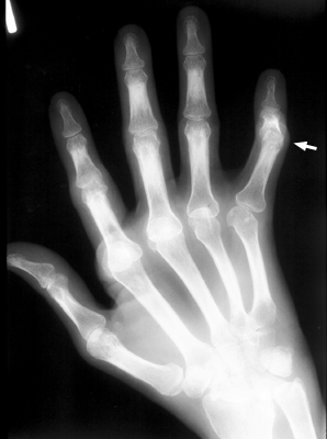
Figure 2 demonstrates the global impact of chronic rheumatoid arthritis across the entire hand and carpus on a posteroanterior (PA) projection. Multiple digits exhibit classic inflammatory sequelae, including marked boutonniere deformities with prominent PIP joint destruction and periarticular demineralization. Concomitant metacarpophalangeal (MCP) joint subluxation, ulnar drift, carpal height loss, and radiocarpal joint space obliteration reflect systemic synovial proliferation. Proliferative synovial pannus within the dorsal PIP capsule undermines the extensor central slip and attenuates the triangular ligament, creating the anatomical substrate for progressive extensor hood failure.
Pathophysiology & Biomechanical Mechanism
The pathophysiology of the boutonniere deformity is rooted in the intricate functional anatomy and reciprocal balance of the digital extensor apparatus. In extensor Zone III (the dorsal PIP joint) and Zone IV (over the proximal phalanx), the extensor digitorum communis (EDC) tendon trifurcates into a robust central slip and two lateral slips. The central slip crosses the PIP joint to insert directly into the dorsal tubercle at the base of the middle phalanx, functioning as the primary dynamic extensor of the PIP articulation. The two lateral slips diverge and receive contributions from the intrinsic hand muscles (lumbricals and interossei) to form the conjoined lateral bands.
Dorsal stability of the lateral bands over the PIP joint is maintained by the triangular ligament (ligamentum triangulare), a thin, transverse fibrous sheet that spans between the paired lateral bands immediately distal to the central slip insertion. Volarly and laterally, the lateral bands are tethered to the flexor tendon sheath by the transverse retinacular ligaments (TRL). Distal to the PIP joint, the lateral bands converge over the dorsum of the middle phalanx (extensor Zone II) to form the terminal extensor tendon, which crosses the DIP joint (extensor Zone I) to insert into the base of the distal phalanx. A secondary stabilizing structure, the oblique retinacular ligament (ORL) of Landsmeer, originates from the palmar aspect of the proximal phalanx and A2 flexor pulley, traverses palmar to the PIP flexion-extension axis, and inserts dorsally into the lateral terminal extensor tendon at the DIP joint.
Biomechanical and cadaveric investigations demonstrate that a boutonniere deformity cannot occur through isolated transection of the central slip alone. When the central slip is disrupted in isolation, the digit exhibits only an extensor lag at the PIP joint (typically 15 to 20 degrees); the lateral bands remain positioned dorsal to the PIP joint flexion-extension axis because they are tethered dorsally by the intact triangular ligament. The development of a true boutonniere deformity requires an obligate second mechanical event: progressive elongation, attenuation, or traumatic tearing of the dorsal triangular ligament.
Once the triangular ligament is incompetent, the transverse retinacular ligaments exert unopposed palmar traction on the lateral bands. Under the influence of repetitive active PIP joint flexion, the lateral bands migrate volarly. As soon as the lateral bands subluxate palmar to the bicondylar coronal axis of rotation of the PIP joint, their mechanical vector reverses completely. Instead of functioning as joint extensors, the displaced lateral bands become paradoxical flexors of the PIP joint. Every attempt by the patient to actively extend the digit activates the EDC and intrinsic muscles, which pulls the volarly displaced lateral bands and paradoxically forces the PIP joint into greater flexion.
As the head of the proximal phalanx herniates dorsally between the subluxated lateral bands (like a button passing through a buttonhole, hence the French descriptor boutonnière), the distance between the proximal extensor origins and the distal insertion is shortened. All proximal extensor excursion generated by the EDC and intrinsics bypasses the defunctioned central slip and is transferred distally into the terminal extensor tendon. This creates severe, unyielding hyperextension of the DIP joint. Over time, tertiary soft-tissue adaptations freeze the deformity: the transverse retinacular ligaments contract and shorten, permanently anchoring the lateral bands in their subluxated volar position; Landsmeer’s oblique retinacular ligaments contract, preventing passive DIP flexion; and the collateral ligaments and volar plate of the PIP joint develop secondary contractures, transforming an initially dynamic, flexible imbalance into a fixed, rigid joint contracture.
The pathophysiological triggers diverge between acute traumatic and chronic inflammatory etiologies. In acute trauma, mechanical failure occurs either through sudden forced PIP flexion against an active extensor contraction (such as a ball striking the tip of an extended finger), a direct dorsal laceration across Zone III, or a volar PIP joint dislocation that avulses the central slip and ruptures the dorsal capsule. In contrast, in rheumatoid arthritis, the deformity develops insidiously as proliferative synovial pannus expands within the retro-extensor pouch beneath the central slip. Chronic synovial hypertrophy and enzymatic degradation stretch the central slip and triangular ligament from deep to superficial, while concomitant subchondral bone erosions weaken the middle phalanx enthesis and destroy articular cartilage.
Modality-Specific Imaging Findings
Plain Radiography
The standard radiographic examination for suspected boutonniere deformity comprises posteroanterior (PA), true lateral, and oblique views of the affected digit, supplemented by a PA view of the whole hand to evaluate global alignment. A true lateral projection of the isolated digit is mandatory; an oblique projection or lateral view of the superimposed hand obscures the precise PIP joint flexion angle, hides minor dorsal avulsion flecks, and precludes accurate measurement of DIP hyperextension. Key radiographic features include:
- Digital Alignment Aberrations: Demonstrates fixed or dynamic PIP joint flexion (ranging from 15 degrees in early cases to greater than 60 degrees in chronic contractures) coupled with reciprocal DIP joint hyperextension (typically 10 to 30 degrees).
- Dorsal Bony Avulsion Fragments: Visualizes small cortical avulsion fractures from the dorsal lip of the middle phalanx base, representing bony central slip avulsions (bony boutonniere). Fragment displacement, articular surface percentage, and comminution must be meticulously documented.
- Joint Congruity and Subluxation: Identifies volar subluxation of the base of the middle phalanx relative to the proximal phalangeal condyles in advanced chronic cases, as well as dorsal prominence of the proximal phalangeal head.
- Inflammatory and Degenerative Features: In rheumatoid arthritis, radiographs depict symmetric PIP joint space loss, periarticular osteopenia, subchondral cyst formation, and marginal erosions at the bare areas of the middle phalanx base. In chronic post-traumatic deformities, secondary osteoarthritis with dorsal osteophytosis and joint space narrowing may be observed.
Computed Tomography
Multidetector computed tomography (CT) utilizing sub-millimeter axial acquisitions (0.5 mm to 1.0 mm slice thickness) with multiplanar 2D coronal, sagittal, and 3D volume-rendered reconstructions provides exceptional osseous characterization in complex or equivocal presentations. Although soft-tissue tendon pathology is better visualized on ultrasound or MRI, CT is indicated in specific clinical scenarios:
- Articular Avulsion Quantification: Precisely measures the size and articular percentage of middle phalanx dorsal avulsion fractures. Involvement of greater than 20% to 30% of the middle phalanx articular surface or fragment displacement exceeding 2 mm is an indication for open or percutaneous reduction and internal fixation.
- Articular Congruity and Step-Off: Evaluates intra-articular step-off and rotation of avulsed fragments that are obscured by overlapping condyles on plain radiographs.
- Preoperative Structural Templating: In chronic Stage III rheumatoid deformities, CT clearly maps the depth of subchondral erosions, residual cortical bone stock, and intra-articular bony bridging/ankylosis, providing essential anatomic data when planning PIP joint arthrodesis or silicone/surface replacement arthroplasty.
Magnetic Resonance Imaging & Ultrasound
Advanced soft-tissue modalities provide direct visualization of the non-osseous components of the digital extensor apparatus, allowing confirmation of central slip tears prior to the development of permanent joint contracture.
High-Resolution Ultrasound: Musculoskeletal ultrasound performed with high-frequency linear transducers (12 to 18 MHz, or ultra-high-frequency probes up to 22 MHz) is an exceptional, rapid bedside imaging tool. Longitudinal views over the dorsal PIP joint display the normal central slip as a uniform, hyperechoic fibrillar band approximately 1.0 mm in thickness inserting onto the middle phalanx dorsal tubercle. Acute central slip disruption appears as focal hypoechoic or anechoic discontinuity, fiber retraction, or complete gap filled with hematoma. Transverse dynamic imaging during gentle passive finger flexion reveals the lateral bands slipping volarly beneath the mid-axial plane of the proximal phalangeal condyles, confirming triangular ligament disruption. Color and Power Doppler demonstrate active hypervascular synovial pannus within the dorsal retro-extensor recess in patients with inflammatory arthritis.
Magnetic Resonance Imaging (MRI): Utilizing dedicated small-parts surface coils or multichannel finger phased-array coils, MRI offers multiplanar soft-tissue and chondral resolution. T1-weighted, T2-weighted, and proton density (PD) fat-suppressed sequences yield specific findings:
- Central Slip Disruption: Sagittal T2/PD fat-suppressed images demonstrate focal hyperintense fluid signal, contour irregularity, or complete discontinuity at the insertion site of the central slip onto the dorsal base of the middle phalanx.
- Triangular Ligament Incompetence & Lateral Band Subluxation: Axial sequences clearly reveal the attenuation or absence of the thin low-signal triangular ligament and show the paired lateral bands displaced palmar to the coronal midline axis of the PIP joint.
- Enthesitis and Marrow Edema: Fluid-sensitive sequences identify bone marrow edema within the middle phalanx base, representing traumatic traction edema or active inflammatory enthesitis.
- Synovial Proliferation and Chondral Status: Post-gadolinium T1-weighted sequences delineate enhancing synovial pannus expanding the dorsal capsule and retro-extensor space, while 3D gradient-echo or intermediate-weighted images assess the degree of PIP articular cartilage degradation.
Diagnostic Utility, Sensitivity & Specificity
The diagnostic evaluation of boutonniere deformity depends heavily on the chronicity of the lesion. In chronic presentations, resting posture and physical examination are obvious, but in the acute phase (the first 10 to 14 days post-injury), clinical inspection alone is notoriously deceptive: the finger often appears minimally swollen with near-normal resting PIP extension, because the lateral bands have not yet stretched the triangular ligament and continue to extend the middle phalanx from their dorsal position. Relying on passive inspection leads to high rates of missed diagnosis and chronic deformity.
The Clinical Elson Test: First described by Reginald Elson in 1986, this provocative clinical maneuver remains the gold standard for detecting acute, isolated extensor central slip disruption before fixed deformity supervenes. The test is executed by flexing the patient’s PIP joint to 90 degrees over the edge of a table or over the examiner’s index finger, thereby taking the lateral bands off stretch. The patient is instructed to actively extend the PIP joint against the examiner’s manual resistance placed over the dorsum of the middle phalanx:
- Normal / Intact Central Slip: The patient generates a strong, rigid active PIP extension force against resistance. Simultaneously, the DIP joint remains completely loose, flaccid, and passively mobile, because all extensor tension is concentrated at the central slip insertion into the middle phalanx.
- Central Slip Disruption: Active PIP extension against resistance is absent or markedly weak. Crucially, the DIP joint becomes rigidly, forcefully hyperextended, resisting passive flexion, because all proximal extensor effort bypasses the ruptured central slip and is shunted through the lateral bands into the terminal extensor tendon.
- Diagnostic Performance: When properly administered in an awake patient, the Elson test exhibits sensitivity approaching 100% and specificity approaching 100% for complete central slip disruption, dramatically outperforming resting visual inspection and static manual testing.
Modified Boyes Test: This complementary clinical maneuver assesses central slip disruption by holding the PIP joint in full passive extension and asking the patient to actively flex the DIP joint. In an acute central slip tear with proximal retraction, the lateral bands are excessively loose, impairing active DIP flexion.
High-Resolution Musculoskeletal Ultrasound Accuracy: In a prospective diagnostic cohort of 21 digits evaluated against surgical exploration, Westerheide, Failla, van Holsbeeck, and Ceulemans (2003) demonstrated that high-resolution sonography (12 to 15 MHz) achieved a sensitivity of 93%, a specificity of 100%, a positive predictive value of 100%, a negative predictive value of 88%, and an overall diagnostic accuracy of 95% for extensor central slip disruptions. Sonography confirmed its utility as an exceptional non-invasive modality to corroborate equivocal Elson tests, particularly when acute pain, local swelling, or digital anesthesia prevents reliable physical testing.
Plain Radiography Utility: Plain radiographs achieve 100% sensitivity for identifying osseous avulsion fractures at the middle phalanx dorsal tubercle. However, for acute closed soft-tissue central slip disruptions without bony involvement, plain radiographs have a sensitivity of less than 20%, as soft tissues are radiolucent and resting alignment remains preserved early on. In chronic stages, radiographs provide 100% specificity for establishing fixed deformity angles, subluxation, and degenerative or inflammatory joint erosion.
Differential Diagnosis & Radiographic Mimics
Accurate differentiation of boutonniere deformity from other digital extensor disorders, flexor contractures, and congenital conditions is critical to avoid inappropriate splinting or destructive surgical exploration. The key diagnostic mimics and their discriminating features are summarized below:
| Sign / Condition | Distinguishing Radiographic Features | Clinical Presentation | Key Differentiating Test |
|---|---|---|---|
| Classic Boutonniere Deformity | PIP joint flexion with reciprocal DIP joint hyperextension; dorsal avulsion fleck from middle phalanx base; joint space loss and marginal erosions in rheumatoid arthritis | Post-traumatic or inflammatory PIP extensor lag progressing to fixed PIP flexion and DIP hyperextension; difficulty flattening hand or making a tight fist | Positive Elson test (weak PIP extension with rigid DIP hyperextension); ultrasound demonstrates central slip gap and volar lateral band migration |
| Swan-Neck Deformity | PIP joint hyperextension coupled with reciprocal DIP joint flexion (exact reciprocal posture of boutonniere); intact central slip; dorsal subluxation of lateral bands | Common in rheumatoid arthritis, trauma, or intrinsic muscle contracture; finger locking in hyperextension; difficulty initiating PIP flexion | Negative Elson test; lateral radiograph demonstrates PIP hyperextension; positive Bunnel-Littler test for intrinsic tightness; intact central slip on ultrasound |
| Mallet Finger (Extensor Zone I) | Isolated DIP joint flexion deformity (extensor lag) with normal or mildly hyperextended PIP joint; dorsal avulsion fleck at base of distal phalanx | History of sudden axial force striking an extended fingertip; unable to actively extend DIP joint; PIP joint maintains normal active motion | Normal PIP kinematics; radiograph shows distal phalanx avulsion or soft-tissue sag at DIP joint; central slip is fully intact on clinical exam and sonography |
| Pseudoboutonniere Deformity | Fixed PIP joint flexion contracture WITHOUT DIP hyperextension; radiographs frequently show calcification or avulsion of the PIP volar plate | History of PIP joint hyperextension injury resulting in volar plate rupture and secondary scar contracture; PIP lacks full extension, but DIP joint has completely normal motion | Negative Elson test; normal active and passive DIP flexion regardless of PIP position; normal central slip and triangular ligament on dynamic ultrasound |
| Camptodactyly | Non-traumatic, painless congenital PIP flexion contracture without DIP hyperextension; typically affects small (5th) finger bilaterally; normal articular contours | Presents in infancy or early adolescence; gradual progressive flexion deformity of the PIP joint; no history of trauma, pain, or systemic inflammatory disease | Negative Elson test; absence of DIP hyperextension; ultrasound demonstrates intact central slip insertion and anomalous lumbrical or flexor digitorum superficialis anatomy |
| Dupuytren Contracture | Palmar nodular and cord thickening causing progressive MCP and PIP flexion contractures without primary DIP hyperextension; normal joint spaces | Adults (predominantly males >50 years); palmar skin puckering and palpable fascial cords (spiral cords, pretendinous cords); gradual loss of extension | Positive Hueston tabletop test (inability to flatten palm on flat surface); palpable palmar/digital cords; intact dorsal extensor apparatus on imaging |
Clinical Significance & Management Implications
The clinical management of boutonniere deformity is structured according to clinical staging systems (such as the Burton and Nalebuff classifications), which categorize the deformity based on joint flexibility, soft-tissue contracture, and articular preservation:
- Stage I (Mild / Early Flexible): PIP joint demonstrates a mild extensor lag (typically 10 to 15 degrees) that is passively correctable to full 0-degree neutral extension; the DIP joint exhibits mild, flexible hyperextension; plain radiographs confirm normal joint spaces and joint congruity.
- Stage II (Moderate / Fixed Soft-Tissue Contracture): PIP flexion deformity progresses to 30 to 40 degrees with limited or absent passive PIP extension; the DIP joint is hyperextended with marked tightness of Landsmeer’s oblique retinacular ligament (loss of passive DIP flexion when the PIP joint is brought toward extension); lateral bands are contracted volarly; plain radiographs reveal preserved joint space without articular cartilage destruction.
- Stage III (Severe / Fixed Joint Destruction): Fixed PIP flexion contracture exceeds 45 to 60 degrees; rigid DIP hyperextension; severe secondary contractures of the collateral ligaments, volar plate, and transverse retinacular ligaments; plain radiographs demonstrate articular cartilage destruction, subchondral erosions (in rheumatoid arthritis), or secondary degenerative osteoarthrosis.
Conservative Management Protocol (First-Line Standard of Care): Closed acute and subacute central slip disruptions (Stage I and flexible Stage II) should universally be treated with a dedicated conservative splinting regimen. Lin and Strauch (2014) established that continuous PIP extension splinting achieves over 85% good-to-excellent functional outcomes in acute closed injuries, avoiding the high stiffness and scar contracture rates associated with acute surgical exploration:
- Isolated PIP Extension Splinting: A custom thermoplastic circumferential splint, dynamic Capener splint, or static digital gutter splint is applied to maintain the PIP joint in absolute 0-degree neutral extension continuously for 6 consecutive weeks. The splint must never be removed for functional activities during this initial period.
- Crucial DIP Joint Freedom: The splint must strictly end immediately distal to the PIP joint, leaving the DIP joint and the MCP joint completely free. Immobilizing the DIP joint along with the PIP joint is a catastrophic technical error that promotes terminal extensor scarring and permanent digital stiffness.
- Active DIP Flexion Exercises: The patient is instructed to perform 10 to 15 repetitions of active DIP joint flexion hourly while wearing the PIP extension splint. Active contraction of the flexor digitorum profundus (FDP) pulls the conjoined lateral bands distally and dorsally across the middle phalanx. This dynamic action prevents contracture of Landsmeer’s oblique retinacular ligament, stretches the contracted transverse retinacular ligaments, and guides the lateral bands back dorsal to the PIP joint flexion-extension axis.
- Weaning and Progressive Mobilization: At the conclusion of 6 weeks, if active PIP extension is maintained without extensor lag, gradual active PIP flexion exercises are introduced, supported by night splinting for an additional 2 to 4 weeks.
Operative Indications and Surgical Techniques: Surgical intervention is reserved for specific anatomical scenarios and presentations refractory to conservative care:
- Acute Open Lacerations: Direct primary anatomical repair of the central slip to the middle phalanx dorsal tubercle using micro-suture anchors or transosseous non-absorbable sutures, accompanied by meticulous re-approximation of the dorsal triangular ligament.
- Acute Bony Boutonniere (Avulsion Fractures): Open reduction and internal fixation (ORIF) utilizing micro-screws, tension-band wiring, or pull-out wire/suture techniques for middle phalanx dorsal avulsion fragments involving greater than 20% to 30% of the articular surface or demonstrating greater than 2 mm of displacement.
- Chronic Flexible Deformities (Littler-Eaton Reconstruction): Centralization and dorsal redistribution of lateral bands. One lateral band is mobilized, transected proximally, and routed dorsally across the PIP joint to augment the central slip and restore middle phalanx extension, while mitigating excessive extensor force transmitted to the DIP joint.
- Chronic Flexible Deformities (Dolphin / Fowler Terminal Tenotomy): Percutaneous or open transection of the terminal extensor tendon over the middle phalanx. This releases the excessive hyperextension tension at the DIP joint, redirecting the proximal extensor force to the middle phalanx to improve active PIP extension, while preserving active DIP extension through Landsmeer’s intact oblique retinacular ligament.
- Chronic Flexible Deformities (Anatomical Tendon Grafting): Central slip reconstruction utilizing a free tendon graft (e.g., palmaris longus or plantaris slip) looped through the middle phalanx base and sutured to the lateral bands, as described by Heng and Chung (2026).
- Chronic Fixed Deformities with Joint Destruction (Stage III): As emphasized by Souter (1974), soft-tissue reconstruction is futile and strictly contraindicated when severe articular cartilage destruction, subchondral erosions, or rigid intra-articular contractures are present. In these advanced cases, definitive salvage procedures include PIP joint arthrodesis in a position of function (typically 40 degrees for the index, 45 degrees for the middle, and 50 degrees for the ring and small fingers) or silicone/pyrocarbon PIP joint replacement arthroplasty in low-demand rheumatoid hands.
Master Class Pro-Tip
Master Class Pro-Tip: Never mistake an acutely normal-appearing finger for an intact extensor apparatus following blunt hyperflexion trauma to the PIP joint. In the first 10 to 14 days post-injury, patients frequently maintain near-normal resting PIP extension because the intact lateral bands remain momentarily dorsal to the joint axis; relying on passive resting posture guarantees a missed diagnosis. Always perform the Elson test: with the PIP joint flexed to 90 degrees over a table edge, an isolated central slip rupture produces immediate, rigid DIP hyperextension upon active extension effort against resistance. When initiating conservative splinting, enforce the cardinal rule of boutonniere management: immobilize only the PIP joint in absolute neutral extension while leaving the DIP joint completely free. Mandating active DIP flexion exercises hourly while in the splint glides the lateral bands dorsally and prevents irreversible contracture of Landsmeer’s oblique retinacular ligament. Finally, never attempt soft-tissue reconstruction on a stiff PIP joint; full passive joint extension must first be restored through serial casting, as soft-tissue transfers cannot overcome a contracted volar plate or collateral ligaments.
Master Class Radiological Pearl: Dynamic Clinical Testing, PIP Splint Isolation & Soft-Tissue Reconstruction Prerequisites
Evidence-Based Synthesis
| Author & Year | Study Design | Sample Size | Key Findings & Diagnostic Accuracy | Clinical Implication | Level of Evidence | DOI |
|---|---|---|---|---|---|---|
| Elson RA (1986) | Clinical Biomechanical Diagnostic Investigation | N = 42 patients / anatomical specimens | Described the pathognomonic “Elson test” for diagnosing isolated extensor central slip disruption prior to the onset of fixed boutonniere deformity: with the PIP joint flexed 90 degrees over the edge of a table, an intact central slip exerts strong PIP extension against resistance while the DIP remains completely floppy and passive. With central slip rupture, the patient demonstrates weak PIP extension while the DIP becomes rigidly extended due to reciprocal lateral band hyper-tension. | Established the universal clinical gold standard for diagnosing occult acute central slip tears before irreversible volar migration of the lateral bands and fixed joint contracture supervene. | Level V | DOI |
| Westerheide KJ, Failla JM, van Holsbeeck M, Ceulemans RY (2003) | Prospective Diagnostic Accuracy Cohort | N = 21 digits | Evaluated high-resolution musculoskeletal ultrasound (12-15 MHz) against surgical exploration for extensor central slip disruption over the PIP joint. Ultrasound demonstrated 93% sensitivity, 100% specificity, and 95% overall diagnostic accuracy in visualizing complete vs. partial central slip tears and characterizing associated triangular ligament disruption and lateral band subluxation. | Validated high-resolution ultrasound as an exceptional non-invasive dynamic imaging modality to confirm occult or equivocal central slip injuries, particularly in swollen digits where clinical testing is ambiguous. | Level II | DOI |
| Pfanner S, Poggetti A (2025) | Contemporary Comprehensive Review & Treatment Algorithm | Systematic Clinical Synthesis | Detailed the progressive pathophysiological staging (Nalebuff classification) of rheumatoid boutonniere deformity: Stage I (mild PIP extensor lag, passively correctable), Stage II (moderate PIP flexion 30-40 degrees, tight oblique retinacular ligament, correctable with passive DIP flexion), and Stage III (fixed PIP flexion deformity with joint destruction, severe DIP hyperextension, and articular cartilage erosion). Highlighted biologic DMARD management, tenosynovectomy, and reconstruction vs. arthrodesis/arthroplasty criteria. | Provides the definitive modern consensus algorithm aligning medical suppression of inflammatory synovitis with stage-specific surgical interventions to preserve manual dexterity. | Level V | DOI |
| Ferrazzini LA, Giesen T (2026) | Cadaveric Anatomic & Kinematic Investigation | N = 16 cadaveric specimens | Quantified the micro-architecture of the triangular ligament, transverse retinacular ligaments, and central slip insertion into the middle phalanx dorsal tubercle. Demonstrated that acute boutonniere deformity cannot occur through central slip transection alone; concomitant lengthening, tearing, or transverse attenuation of the dorsal triangular ligament is strictly required to permit the lateral bands to subluxate volar to the PIP flexion-extension axis. | Redefined anatomical principles of extensor repair, emphasizing that anatomical reconstruction must reconstitute both central slip length and dorsal transverse tethering of the lateral bands. | Level V | DOI |
| Lin JS, Strauch RJ (2014) | Evidence-Based Clinical & Imaging Review | Systematic Evidence Synthesis | Analyzed conservative splinting outcomes versus primary surgical repair for acute closed central slip disruptions. Established that 6 weeks of continuous PIP joint extension splinting (leaving the DIP and MCP joints completely free for active flexion exercises to glide the lateral bands) yields over 85% good-to-excellent functional results, whereas surgical repair of closed acute tears has higher rates of stiffness and scar contracture. | Confirmed continuous PIP extension splinting with unrestricted active DIP flexion as the first-line evidence-based standard of care for acute closed boutonniere injuries. | Level IV | DOI |
| Heng SY, Chung SR (2026) | Surgical Technique & Clinical Series | N = 18 patients | Outlined refined surgical reconstruction for chronic, flexible Stage II boutonniere deformities using lateral band re-routing combined with terminal extensor tenotomy (Dolphin/Fowler principle). Achieved an average active PIP extension improvement of 38 degrees and restored functional composite finger flexion without recurrent extensor lag. | Demonstrates reliable operative correction of chronic post-traumatic and early inflammatory boutonniere deformities refractory to conservative splinting. | Level IV | DOI |
| Littler JW, Eaton RG (1967) | Landmark Biomechanical & Surgical Cohort Study | N = 31 digits | Formulated the foundational biomechanical concept of vector redistribution in the extensor hood: transection of one lateral band, mobilization, and dorsal translocation across the PIP joint to re-establish middle phalanx extension while simultaneously mitigating excessive hyperextension forces at the DIP joint. | Provided the classic surgical blueprint for reconstructing soft-tissue extensor balance in chronic flexible boutonniere deformity. | Level IV | DOI |
| Souter WA (1974) | Comprehensive Clinical Review & Multi-Center Analysis | N = 106 digits | Critical evaluation of surgical vs. non-operative treatments for post-traumatic and rheumatoid boutonniere deformities; established that chronic fixed contractures respond poorly to soft-tissue tendon balancing alone if joint cartilage destruction or severe collateral ligament shortening is present, advocating joint fusion in advanced destruction. | Underscored that operative success strictly depends on whether the PIP joint can be passively brought to neutral extension prior to extensor reconstruction. | Level IV | DOI |
| Massengill JB (1992) | Comprehensive Narrative Review & Classification | Narrative Synthesis | Synthesized the natural history of boutonniere deformity from acute closed trauma, open lacerations, and chronic rheumatoid proliferative synovitis; emphasized the dual pathology of PIP central slip elongation/attenuation and volar migration of lateral bands with contracture of the transverse retinacular ligaments and oblique retinacular ligaments. | Defined the classic diagnostic criteria and sequential stages of extensor mechanism imbalance cited across hand surgery curricula. | Level V | DOI |
| Rizio L, Belsky MR (1996) | Clinical Review & Pathogenetic Analysis | Narrative Clinical Review | Elucidated the pathogenetic differences between boutonniere and swan-neck deformities in rheumatoid arthritis, illustrating how PIP synovitis distends the dorsal capsule and stretches the central slip, whereas MCP synovitis precipitates intrinsic tightness favoring swan-neck deformity. | Outlined precise differential diagnostic physical exam maneuvers and articulated guidelines for timing of surgical synovectomy to arrest extensor apparatus breakdown. | Level V | DOI |
References
- Elson RA. Rupture of the central slip of the extensor hood of the finger. A test for early diagnosis. J Bone Joint Surg Br. 1986;68(2):229-231. DOI [Level V: Clinical Biomechanical Diagnostic Investigation]
- Westerheide KJ, Failla JM, van Holsbeeck M, Ceulemans RY. Ultrasound visualization of central slip injuries of the finger extensor mechanism. J Hand Surg Am. 2003;28(6):1009-1013. DOI [Level II: Prospective Diagnostic Accuracy Cohort]
- Pfanner S, Poggetti A. Swan-Neck and Boutonniere Deformity in Rheumatoid Hand. Hand Clin. 2025;41(1):47-58. DOI [Level V: Contemporary Comprehensive Review & Treatment Algorithm]
- Ferrazzini LA, Giesen T. New perspective on acute boutonniere deformity: An anatomical study with illustrative case report. J Hand Microsurg. 2026;18(3):100490. DOI [Level V: Cadaveric Anatomic & Kinematic Investigation]
- Lin JS, Strauch RJ. Closed Soft Tissue Extensor Mechanism Injuries (Mallet, Boutonniere, and Sagittal Band). J Hand Surg Am. 2014;39(5):1005-1011. DOI [Level IV: Evidence-Based Clinical & Imaging Review]
- Heng SY, Chung SR. Technique for Central Slip Reconstruction for Chronic Boutonniere Deformity. Tech Hand Up Extrem Surg. 2026;30(2):e0551. DOI [Level IV: Surgical Technique & Clinical Series]
- Littler JW, Eaton RG. Redistribution of Forces in the Correction of the Boutonnière Deformity. J Bone Joint Surg Am. 1967;49(7):1267-1274. DOI [Level IV: Landmark Biomechanical & Surgical Cohort Study]
- Souter WA. The Problem of Boutonniere Deformity. Clin Orthop Relat Res. 1974;(104):116-133. DOI [Level IV: Comprehensive Clinical Review & Multi-Center Analysis]
- Massengill JB. The Boutonniere Deformity. Hand Clin. 1992;8(4):787-801. DOI [Level V: Comprehensive Narrative Review & Classification]
- Rizio L, Belsky MR. Finger deformities in rheumatoid arthritis. Hand Clin. 1996;12(3):531-540. DOI [Level V: Clinical Review & Pathogenetic Analysis]
Last Updated on September 29, 2026
🔗 Knowledge Graph & Clinical Correlates
OrthopaedicsOne Living Semantic Network🛠️ Operative Techniques
- Wrist Implant Arthroplasty Hand and Wrist
- Wrist disarticulation Hand and Wrist
- Wrist Dennervation Hand and Wrist
- Wrist arthroscopy Hand and Wrist
🩺 Clinical Conditions & Indications
- Wrist sprains 1. High-Yield Summary Wrist sprain is an imprecise clinical descriptor that frequently obscures high-energy ligamentous disruption of the...
- Wartenberg’s syndrome 1. High-Yield Summary Wartenberg's syndrome (cheiralgia paresthetica) is an entrapment neuropathy of the superficial branch of the radial...
- Volar plate injury 1. High-Yield Summary Volar plate injuries of the proximal interphalangeal (PIP) joint represent the most frequent soft-tissue and...
- Viral arthritis 1. High-Yield Summary Viral arthritis is an acute, systemic infectious arthropathy characterized by transient or persistent synovial inflammation...