Arthroscopic Treatment of Lateral Epicondylitis
Since the original description of lateral epicondylitis by Runge in 1873, there has been controversy regarding the etiology, pathoanatomy and most appropriate methods of nonoperative and operative management. Although commonly referred to as “tennis elbow” because of an initial association with lawn tennis, lateral epicondylitis can be caused by minor trauma or overuse with repetitive motion of the wrist while the arm is extended. The accepted theory of the pathogenesis of lateral epicondylitis is that overuse leads to microscopic tears in the origin of the extensor carpi radialis brevis (ECRB), with subsequent tendon replacement by immature reparative tissue consisting of disorganized collagen, fibroblasts, and vascular elements (angiofibroblastic tendinosis).1-3
Most patients respond to nonoperative management, such as activity modification, bracing, and steroid injections. Low-cost counterforce braces may transfer force from the ECRB and its origin and distribute it more evenly, allowing the tendon to heal. Steroid injections may enhance early recovery but can cause calcium deposits, and recent studies have not shown consistent long-term benefits.
Between 4% and 11% of patients fail nonoperative management of lateral epicondylitis and require operative treatment for disabling, recalcitrant symptoms.1,4-6 Traditionally, the standard procedure for lateral epicondylitis has been an open release as described by Nirschl. However, outcomes of arthroscopic techniques have shown to be as reliable but with several advantages.7-15
 Read more…
Surgery is indicated for patients who fail a comprehensive course of nonoperative treatment and have continued pain that interferes with daily activities. Most physicians accept a 6-month course of nonoperative treatment. Before surgery, it is important to assess compliance and motivation, which may explain why a patient failed to respond to nonoperative treatment.
Technique
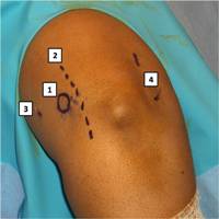
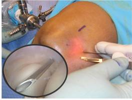
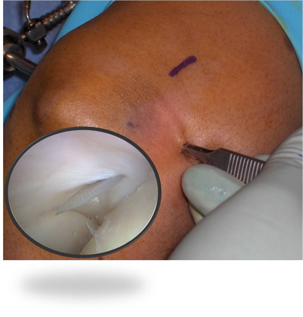
The patient is positioned in the lateral decubitus position with the arm over an elbow holder. A proximal anteromedial portal is created approximately 2 cm proximal to the medial epicondyle and 1 cm anterior to the medial intermuscular septum (Figure 1). A 4.0-mm, 30-degree arthroscope is inserted over a switching stick, and from the proximal anteromedial portal, the radiocapitellar articulation and adjacent joint capsule are examined. Pronation and supination of the forearm allow for full examination of the radial head. An 18-gauge needle is used to localize the anterolateral portal using an outside-in technique, and a 15 blade is used to create the portal and release the majority of the ECRB tendon off the lateral epicondyle under direct visualization (Figure 2 and Figure 3). A 4.0-mm shaver is inserted and the capsule surrounding the portal is debrided and removed (Figure 4).
Figure 1. Anatomic landmarks for arthroscopic lateral release. 1 – Medial epicondyle, 2 – Ulnar nerve, 3 – Proximal anteromedial portal, 4 – Radiocapitellar joint.
Figure 2. Needle localization of radiocapitellar joint and creation of anterolateral portal using outside-in technique.
Figure 3. Creation of anterolateral portal and release of ECRB under direct visualization.
Figure 4. Debridement of capsule and pathologic ECRB tendon origin with shaver.
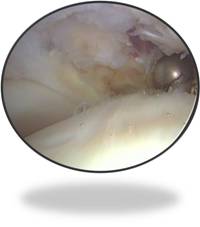
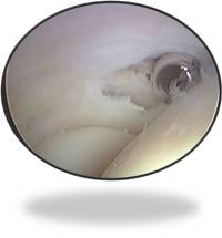
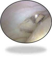
The diseased ECRB tendon lies between the capsule and the overlying extensor digitorum communis. An Arthrocare bipolar probe can be used to release the remainder of the ECRB from the lateral epicondyle (Figure 5). The release is carried distally, making certain to remain above the equator of the radial head to prevent injury to the lateral collateral ligament. Debridement of the ECRB is considered complete when all visibly abnormal tissue has been removed (Figure 6). Portals are closed with a 3-0 prolene suture.
Figure 5. Completion of debridement with Arthrocare ablation.
Figure 6. Debridement of the ECRB is considered complete when all visibly abnormal tissue has been removed.
Advantages of Arthroscopic Over Open Lateral Release
- Smaller incisions, minimal morbidity to the soft tissues and thus less postoperative pain. Although anecdotal, surgeons who have performed both open and arthroscopic lateral releases have noticed improved pain levels in the day surgery unit and subsequent follow-up after arthroscopic techniques.
- Ability to address coexistent intra-articular pathology (synovitis, plica). Several authors using arthroscopic techniques have noted associated intra-articular lesions present in 19% to 44% of patients.10,13,15 Baker et al reported a higher incidence of intra-articular lesions. In their early experience, they routinely looked into the posterior compartment of the elbow, which they no longer do. Nirschl and Pettrone noted an 11% incidence of associated intra-articular disorders using an open approach. Failure to address associated intra-articular lesions may be a reason for persistent symptoms despite adequate open release.
- Only the tendon affected by tennis elbow (ECRB) is released in the arthroscopic method, as this tendon is the closest extensor tendon to the elbow joint. With the open procedure, other extensor tendons are split or released, leading to increased soft tissue morbidity and weak grip strength postop. Several cadaveric studies have demonstrated the efficacy of arthroscopic techniques in removing 100% of the ECRB origin without violating the lateral ulnar collateral ligament or injury to adjacent neurovascular structures.16,17 Cummins recently assessed the effectiveness of the arthroscopic debridement. After an initial arthroscopic debridement, a traditional open exposure was performed with gross and histological analysis done. The results suggest that after an initial learning curve, arthroscopic debridement can effectively remove all of the gross pathologic tissue seen during a traditional open exposure.
- Accelerated rehabilitation and earlier return to sports and work with no loss of grip strength. Baker et al reported a mean return-to-work interval of 2.2 weeks. Owens et al13 reported a mean return to work of 6 days in their military population, while Jerosch et al10 showed that their patients who were primarily tradesmen and office workers returned to work at a mean of 3.2 weeks. Although open procedures achieve a high level of satisfactory results, they typically have a longer recovery period. In the study by Nirschl and Pettrone, patients with good to excellent results took a mean of 2.6 months to be symptom free. Verhaar et al18 reported that only eight of 44 patients returned to work by 6 weeks after open release; at 12 weeks, 13 patients were still unable to return to work.
There are no prospective, randomized studies in the literature comparing arthroscopic debridement of the ECRB origin with open release, and there are only a few retrospective, non-randomized studies comparing arthroscopic release with other operative treatments. Szabo et al15 compared patients who underwent a lateral release done percutaneously, arthroscopically, or open with a mean follow-up of 48 months. There were no significant differences with respect to complications, failures, pain scores, or outcome scores. Peart et al14 reported on 46 patients with open releases compared to 29 patients with arthroscopic releases. Patients treated with arthroscopic release returned to work sooner and required less postoperative therapy. Baker et al compared 15 open Nirschl procedures with 15 arthroscopic releases in a non-randomized trial and found a significant difference in the return-to-work and sports rate, with a mean of 66 days in the open group and 35 days in the arthroscopic group.
Several authors have published successful results at short-term follow-up using similar arthroscopic techniques with minor modifications.10,13-15 Baker et al have shown that these short-term results are maintained in the long-term. They retrospectively reviewed 30 patients who had undergone arthroscopic lateral release, with a mean follow-up of 11 years. The average pain score at rest was 0; with activities of daily living, 1.0; and with work or sports, 1.9. The mean functional score was 11.7 out of a possible 12 points. No patient required further surgery or repeat injections after surgery. One patient continued to wear a counterforce brace with heavy activities. Twenty-three patients (77%) stated they were “much better,” six patients (20%) stated they were “better,” and one patient (3%) stated he was the same. Twenty-six patients (87%) were satisfied, and 28 patients (93%) stated they would have the surgery again if needed.
Conclusion
High rates of good to excellent results have been reported regardless of the type of surgical technique used for the operative treatment of lateral epicondylitis. There is a lack of controlled trials to support one technique over another. However, the current literature suggests that arthroscopic lateral release results in less soft tissue morbidity, improved ability to address co-existent pathology, and accelerated rehabilitation with earlier return to sports and work as compared with open techniques. In addition, short- and long-term outcomes are equivalent or better, with no increased rate of complications.
Reprinted with permission from the Fall 2008 issue of COA Bulletin
Additional Reading
Runge F. Zur genese und behandlung des schreibekrampfes. Berl Klin Wochenschr 1873;10:245-248
References
- Nirschl RP, 1992. “Elbow tendinosis/tennis elbow.” Clin Sports Med 11 (4): 851-70
- Nirschl RP, Pettrone FA, 1979. “Tennis elbow. The surgical treatment of lateral epicondylitis.” J Bone Joint Surg Am 61 (6A): 832-9
- Lo MY, Safran MR, 2007. “Surgical treatment of lateral epicondylitis: a systematic review.” Clin Orthop Relat Res 463: 98-106
- Bowen RE, Dorey FJ, Shapiro MS, 2001. “Efficacy of nonoperative treatment for lateral epicondylitis.” Am J Orthop (Belle Mead NJ) 30 (8): 642-6
- Boyd HB, McLeod AC, 1973. “Tennis elbow.” J Bone Joint Surg Am 55 (6): 1183-7
- Coonrad RW, Hooper WR, 1973. “Tennis elbow: its course, natural history, conservative and surgical management.” J Bone Joint Surg Am 55 (6): 1177-82
- Baker CL, Murphy KP, Gottlob CA, Curd DT, 2000 Nov-Dec. “Arthroscopic classification and treatment of lateral epicondylitis: two-year clinical results.” J Shoulder Elbow Surg 9 (6): 475-82
- Baker CL, Baker CL, 2008. “Long-term follow-up of arthroscopic treatment of lateral epicondylitis.” Am J Sports Med 36 (2): 254-60
- Cummins CA, 2006. “Lateral epicondylitis: in vivo assessment of arthroscopic debridement and correlation with patient outcomes.” Am J Sports Med 34 (9): 1486-91
- Jerosch J, Schunck J, 2006. “Arthroscopic treatment of lateral epicondylitis: indication, technique and early results.” Knee Surg Sports Traumatol Arthrosc 14 (4): 379-82
- Kuklo TR, Taylor KF, Murphy KP, Islinger RB, Heekin RD, Baker CL, 1999. “Arthroscopic release for lateral epicondylitis: a cadaveric model.” Arthroscopy 15 (3): 259-64
- Mullett H, Sprague M, Brown G, Hausman M, 2005. “Arthroscopic treatment of lateral epicondylitis: clinical and cadaveric studies.” Clin Orthop Relat Res 439: 123-8
- Owens BD, Murphy KP, Kuklo TR, 2001. “Arthroscopic release for lateral epicondylitis.” Arthroscopy 17 (6): 582-7
- Peart RE, Strickler SS, Schweitzer KM, 2004. “Lateral epicondylitis: a comparative study of open and arthroscopic lateral release.” Am J Orthop (Belle Mead NJ) 33 (11): 565-7
- Szabo SJ, Savoie FH, Field LD, Ramsey JR, Hosemann CD, 2006 Nov-Dec. “Tendinosis of the extensor carpi radialis brevis: an evaluation of three methods of operative treatment.” J Shoulder Elbow Surg 15 (6): 721-7
- Cohen MS, Romeo AA, Hennigan SP, Gordon M, 2008 Nov-Dec. “Lateral epicondylitis: anatomic relationships of the extensor tendon origins and implications for arthroscopic treatment.” J Shoulder Elbow Surg 17 (6): 954-60
- Smith AM, Castle JA, Ruch DS, 2003 Jul-Aug. “Arthroscopic resection of the common extensor origin: anatomic considerations.” J Shoulder Elbow Surg 12 (4): 375-9
- Verhaar J, Walenkamp G, Kester A, van Mameren H, van der Linden T, 1993. “Lateral extensor release for tennis elbow. A prospective long-term follow-up study.” J Bone Joint Surg Am 75 (7): 1034-43
Attachments:

Lateral_Epi_Arthro_Figure_1.jpg (image/jpeg)
Lateral_Epi_Arthro_Figure_2.jpg (image/jpeg)
Lateral_Epi_Arthro_Figure_3.jpg (image/jpeg)
Lateral_Epi_Arthro_Figure_4.jpg (image/jpeg)
Lateral_Epi_Arthro_Figure_5.jpg (image/jpeg)
Lateral_Epi_Arthro_Figure_6.jpg (image/jpeg)